
FIGURE
Fig. 3
- ID
- ZDB-FIG-070911-7
- Publication
- Perner et al., 2007 - The Wilms tumor genes wt1a and wt1b control different steps during formation of the zebrafish pronephros
- Other Figures
- All Figure Page
- Back to All Figure Page
Fig. 3
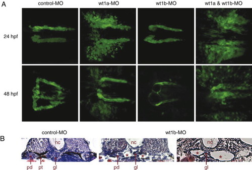
Knockdown of wt1a and wt1b leads to different defects during pronephros development. (A) Confocal images of control and wt1 morpholino-injected wt1b::GFP embryos were recorded 24 and 48 h after fertilization and assembled into 3D projections. (B) Formation of kidney cysts (asterisks) in wt1b morpholino-injected embryos 48 h after fertilization was visualized by H&E staining on cross sections. Note that a part of the glomerular filtration unit was still detectable between the cystic structures. nc, notochord; gl, glomerulus; pt, pronephric tubule; pd, pronephric duct. |
Expression Data
Expression Detail
Antibody Labeling
Phenotype Data
| Fish: | |
|---|---|
| Knockdown Reagents: | |
| Observed In: | |
| Stage Range: | Prim-5 to Long-pec |
Phenotype Detail
Acknowledgments
This image is the copyrighted work of the attributed author or publisher, and
ZFIN has permission only to display this image to its users.
Additional permissions should be obtained from the applicable author or publisher of the image.
Reprinted from Developmental Biology, 309(1), Perner, B., Englert, C., and Bollig, F., The Wilms tumor genes wt1a and wt1b control different steps during formation of the zebrafish pronephros, 87-96, Copyright (2007) with permission from Elsevier. Full text @ Dev. Biol.