FIGURE SUMMARY
Title

The Glutamine Synthetases Are Required for Sensory Hair Cell Formation and Auditory Function in Zebrafish

Authors
Zhao, Y., Wang, Z., Xu, M., Qian, F., Wei, G., Liu, D.
Source
Full text @ Int. J. Mol. Sci.

ZFIN is incorporating published figure images and captions as part of an ongoing project. Figures from some publications have not yet been curated, or are not available for display because of copyright restrictions.

PHENOTYPE:
Fish:
Condition:
Knockdown Reagent:
Observed In:
Stage: Days 7-13

ZFIN is incorporating published figure images and captions as part of an ongoing project. Figures from some publications have not yet been curated, or are not available for display because of copyright restrictions.

Phylogenetic analysis of Glul across diverse species and the expression patterns of the Glul gene family in zebrafish. (A) The MEGA software (Version 11.0.13) was utilized to construct a phylogenetic tree based on amino acid sequences, encompassing a diverse range of species including H. sapiens, P. troglodytes, M. mulatta, C. lupus, B. taurus, M. musculus, R. norvegicus, G. gallus, D. rerio, D. melanogaster, A. gambiae, C. elegans, S. cerevisiae, K. lactis, E. gossypii, S. pombe, N. crassa, A. thaliana, O. sativa, and X. tropicalis. Branches of the same color are closer. Whole-mount in situ hybridization (WISH) was performed using glula, glulb, and glulc probes in wild-type zebrafish. (B) The results demonstrated that the expression of the glula gene was observed specifically in the otic vesicle. Green arrowheads indicate otic vesicle. Boxes indicate the magnified regions. (C) while the glulb gene exhibited expression in both the otic vesicle and lateral line. Green arrowheads indicate otic vesicle and neuromasts. Boxes indicate the magnified regions. (D) Conversely, no significant signal was detected for the glulc gene.

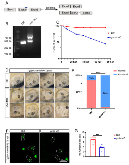
The downregulation of glula results in a reduced survival rate and impacts otolith development. (A) Diagram illustrating the glula-MO binding site and potential cleavage methods. (B) Reproducible knockdown of the glula gene was successfully demonstrated through PCR experimentation. (C) Knockdown of glula leads to a significant decrease in the survival rate of zebrafish. (D) Abnormal otolith development was observed in zebrafish following glula-MO injection. (E) Statistical analysis revealed that 98% of zebrafish injected with glula-MO exhibited aberrant otolith development. Chi-squared test: ****, p < 0.0001. (F) After the injection of glula-MO, a significant decrease in hair cells was observed in otic vesicles. Dash line cycles indicate the crista hair cells. (G) Statistical analysis of the hair cell number in otic vesicles. t-test: ***, p < 0.001.

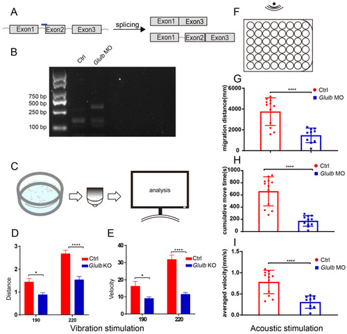
The knockdown of glulb expression resulted in a delayed response of zebrafish to both vibrational and auditory stimuli. (A) Schematic representation of the glulb-MO binding site and potential cleavage methodologies. (B) Successful glulb gene knockdown validated by PCR analysis. (C) Diagram illustrating the experimental setup for vibration stimulation: 7-day-old juvenile zebrafish were positioned within the mold, exposed to vibration stimulation, and subsequently subjected to data recording and analysis. (D,E) Statistical analysis revealed a significant decrease in the distance traveled and swimming velocity of zebrafish in the glulb-MO group compared to the Ctrl group. t-test: *, p < 0.05; ****, p < 0.0001. (F) Five-day-old zebrafish juveniles were placed in the orifice plate, exposed to acoustic stimulus, and subjected to subsequent data recording and analysis. (GI) The glulb-MO group exhibited a significant reduction in the movement distance, average speed, and sustained movement time of zebrafish compared to the control group, as revealed by statistical analysis. t-test: ****, p < 0.0001.

PHENOTYPE:
Fish:
Conditions:
Knockdown Reagent:
Observed In:
Stage Range: Day 5 to Days 7-13

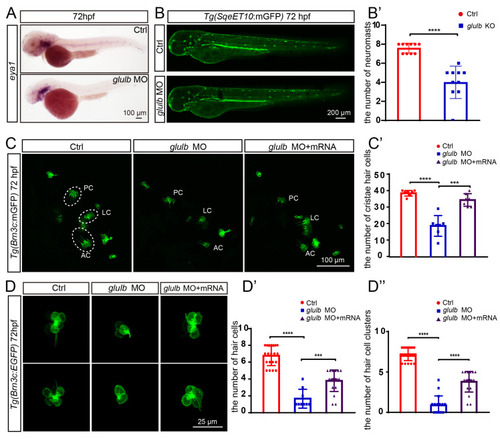
The zebrafish that were injected with glulb-MO exhibited a significant decrease in the number of neuromasts, hair cell clusters, and hair cells, and co-injection of glulb mRNA successfully rescued the observed phenotype following glulb knockdown. (A) In situ hybridization using the eya1 probe revealed a reduced number of neuromasts in the glulb-MO zebrafish group compared to the Ctrl group at 72 hpf. (B) The application of laser confocal microscopy revealed a decrease in the number of neuromasts in glulb-MO zebrafish embryos at 72 hpf compared to the control zebrafish, and (B’) the observed discrepancy exhibited statistical significance. t-test: ****, p < 0.0001. (C) The number of hair cells in the inner ear of glulb-MO zebrafish exhibited a significant decrease compared to the control group. Co-injection of glulb mRNA effectively rescued this phenotype, and (C’) subsequent statistical analysis revealed statistically significant differences. One-way ANOVA: ****, p < 0.0001; ***, p < 0.001. (D) The glulb-MO zebrafish exhibited decreased hair cells within the L3 neuromasts, which can be rescued by co-injection of glulb mRNA. (D’) Statistical analysis revealed a significant difference. (D”) A statistical analysis of hair cell clusters in the control, glulb-MO injected, and glulb-MO co-injection with glulb mRNA embryos was conducted. One-way ANOVA: ****, p < 0.0001; ***, p < 0.001.

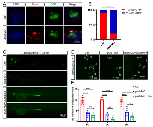
Knockdown of glulb induces apoptosis in zebrafish hair cells, and the supplementation of glutamine fails to rescue the observed phenotype. (A) The zebrafish embryos treated with glulb-MO and the control zebrafish were subjected to TUNEL staining, which revealed the presence of apoptotic signals, specifically in the hair cells of the glulb-MO-treated zebrafish. (B) The percentage of hair cells displaying apoptotic signals was evaluated in control zebrafish embryos and glulb-MO-injected zebrafish. TUNEL+/GFP+ denotes hair cells exhibiting apoptotic signals, while TUNEL-/GFP+ represents hair cells lacking apoptotic signals. Chi-squared test: ****, p < 0.0001. (C) The phenotype of hair cell clusters in whole embryonic zebrafish was visualized using confocal laser microscopy. Significantly reduced hair cell clusters were observed in glulb-MO-treated zebrafish compared to Ctrl zebrafish, and co-injection of glulb-MO and glutamine failed to rescue the reduction in hair cell clusters. (D) Similarly, a decreased number of hair cells was observed in the inner ear of zebrafish embryos treated with glulb-MO compared to control zebrafish, and co-injection of glulb-MO and glutamine failed to rescue the phenotype. Lateral crista (LC), anterior crista (AC) and posterior crista (PC). (E) The number of hair cells exhibited a significant reduction statistically, with no substantial rescue observed following glutamine supplementation. One-way ANOVA: ****, p < 0.0001; **, p < 0.01, ns means no significant.

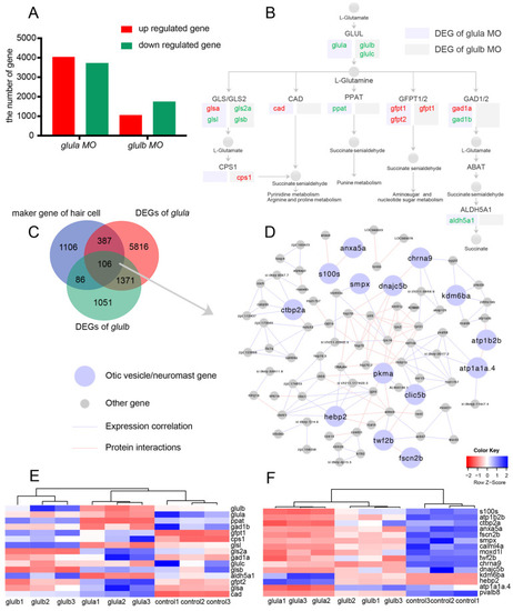
Transcriptomic analysis of zebrafish with glula knockdown and glulb knockdown. (A) Differentially expressed genes (DEGs) in the transcriptome of glula knockdown zebrafish and glulb knockdown zebrafish were identified. (B) DEGs in the glutamine pathway were observed in the transcriptomes of glula knockdown and glulb knockdown zebrafish. (C) The overlap of DEGs in glula transcriptomes, glulb transcriptomes, and the hair cell marker genes was identified through single-cell sequencing. (D) In the gene interaction network of overlap DEGs, pink nodes represent gene expression in the neuromasts, purple nodes indicate gene expression in the otic vesicles, gray nodes denote other genes, purple edges signify gene expression correlation, and pink edges indicate protein interactions. (E) Heatmaps illustrating DEGs in the glul pathway. (F) Heatmaps of DEGs in neuromasts and otic vesicles within the gene–gene interaction network.

Acknowledgments
This image is the copyrighted work of the attributed author or publisher, and ZFIN has permission only to display this image to its users. Additional permissions should be obtained from the applicable author or publisher of the image. Full text @ Int. J. Mol. Sci.