FIGURE SUMMARY
Title

Presynaptic Nrxn3 is essential for ribbon-synapse maturation in hair cells

Authors
Jukic, A., Lei, Z., Cebul, E.R., Pinter, K., Tadesse, Y., Jarysta, A., David, S., Mosqueda, N., Tarchini, B., Kindt, K.
Source
Full text @ Development

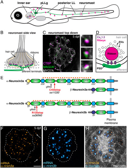
α-nrxn3a and α-nrxn3b are expressed in lateral-line hair cells. (A) Diagram of a 5 day post fertilization (dpf) larval zebrafish showing sensory hair-cell clusters in the inner ear and posterior lateral line (neuromasts). Neurons from the posterior lateral line ganglia (pLLg) innervate (pLLg, green) lateral-line neuromasts. (B) Side view of a neuromast organ showing hair cells, presynapses (ribbons) and afferent processes. The dashed box outlines the synaptic layer. (C) Immunostaining of the synaptic layer, viewed from above. CTBP and MAGUK stain presynapses/ribbons (magenta) and postsynapses (green), respectively. MYO7A stains hair cells (gray). Higher magnification of three synapses is shown on the right. (D) Diagram of a hair-cell ribbon synapse. The presynapse/ribbon consists mainly of Ribeye, a splice variant of CTBP2. The ribbon is surrounded by glutamate-filled synaptic vesicles (SVs). CaV1.3 channels cluster beneath the ribbon, opposite the postsynaptic density (PSD). (E) Zebrafish have two orthologues of Nrxn3, Nrxn3a and Nrxn3b. Each neurexin has a long α form and a shorter β form. Red arrows show location of the mutations in germline zebrafish mutants; these lesions disrupt an obligatory exon in the α form of each orthologue (C455stop and R134stop). Location of gRNAs used in a crispant analysis are indicated. The α and β forms each have a unique start and signal peptide (SP). Each α form has six Laminin G-like domains (LNS) and three epidermal growth factor-like domains (EGF). The red dashed line indicates the location of the RNA FISH probes used in F-H. (F-H) RNA FISH analysis reveals that both α-nrxn3a (F, orange) and α-nrxn3b (G, cyan) mRNAs are present in lateral-line hair cells. Arrows in F indicate α-nrxn3a puncta that are present outside of hair cells, likely in supporting cells. In H, hair cells (myo6b:memGCaMP6 s) are shown in grayscale. The dashed lines in F-H outline the location of hair cells. All images are from larvae at 5 dpf. Scale bars: 5 µm (C,F): 1 µm (C, inset).

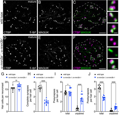
Loss of α-Nrxn3 impairs synapse organization in mature lateral-line hair cells. (A-F) Confocal images of mature neuromasts (5 dpf) from wild-type controls (A-C) and α-nrxn3a; α-nrxn3b mutants (D-F). CTBP labels presynapses (A,D) and MAGUK labels postsynapses (B,E). Merged images are shown in C and F. Dashed lines in C and F outline the hair-cell region [via MYO7A co-label (not shown)]. The boxed areas (magnified on right) in C and F show three examples of individual synapses. (G-J) Quantification shows a similar number of hair cells per neuromast (G), but significantly fewer complete synapses per hair cell in α-nrxn3a; α-nrxn3b mutants (H). There are more unpaired presynapses (I) and postsynapses (J) per hair cell in α-nrxn3a; α-nrxn3b. The total number of presynapses remains unchanged (I), whereas the total number of postsynapses (J) per hair cell is decreased in nrxn3a; nrxn3b mutants. n=10 wild-type and 11 α-nrxn3a; α-nrxn3b mutant neuromasts at 5 dpf. An unpaired two-tailed t-test was used in G and H, and a two-way ANOVA was used in I and J. ns, P>0.05; ****P<0.0001. Data are mean±s.e.m. Scale bars: 5 µm (C,F); 0.5 µm (C,F, insets).

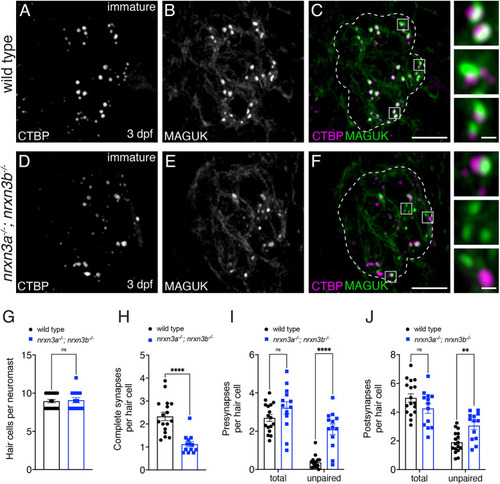
Nrxn3 is required for early synapse maturation in lateral-line hair cells. (A-F) Confocal images of developing hair cells (3 dpf) from wild-type controls (A-C) and nrxn3a; nrxn3b mutants (D-F). CTBP labels presynapses (A,D) and MAGUK labels postsynapses (B,E). Merged images are shown in C and F. The boxed areas (magnified on right) in C and F show three examples of individual synapses. Dashed lines in C and F outline the hair-cell region [via a MYO7A co-label (not shown)]. (G-J) Quantification shows a similar number of hair cells per neuromast (G), but significantly fewer complete synapses per hair cell in nrxn3a; nrxn3b mutants (H). There are significantly more unpaired presynapses (I) and postsynapses (J) per hair cell in nrxn3a; nrxn3b mutants. In developing hair cells, there is no change in the total number of presynapses (I) or postsynapses (J) per hair cell in nrxn3a; nrxn3b mutants. n=17 wild-type and 13 nrxn3a; nrxn3b mutant neuromasts. An unpaired two-tailed t-test was used in G and H, and a two-way ANOVA was used in I and J. ns, P>0.05; **P<0.01; ****P<0.0001. Data are mean±s.e.m. Scale bars: 5 µm (C,F); 0.5 µm (C,F, insets).

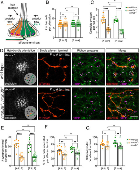
Nrxn3 impacts synapse loss and selectivity to a greater extent in hair cells sensing anterior flow. (A) Primary pLL neuromasts have two populations of hair cells: one responds to anterior flow (green, P to A) and the other responds to posterior flow (orange, A to P). Each population is selectively innervated by distinct afferent neurons (green and orange terminals). (B) Phalloidin label reveals similar numbers of hair cells per neuromast oriented P-to-A and A-to-P flow in wild type and nrxn3a; nrxn3b mutants. n=21 wild-type and 23 nrxn3a; nrxn3b mutant neuromasts at 5 dpf. (C) There are fewer complete synapses in hair cells responding to P-to-A and A-to-P flow in nrxn3a; nrxn3b mutants. There is also a significant reduction in complete synapses in hair cells responding to P to A flow compared with those responding to A to P flow in nrxn3a; nrxn3b mutants. n=10 wild-type and 11 nrxn3a; nrxn3b mutant neuromasts at 5 dpf. (D) Single afferent terminal labeling (P to A fibers) in wild type (top) and an nrxn3a; nrxn3b mutant (bottom). Phalloidin labels hair bundles (orientation indicated by arrows); tdTomato labels individual afferent terminals; CTBP labels presynapses; MAGUK labels postsynapses, and faintly labels hair-cell outlines to provide context. The green and orange arrows in the Phalloidin images indicate the orientation of hair cells that are correctly or incorrectly innervated by each terminal, respectively. The black arrows indicate hair cells that are not innervated by the terminal. In the merged images, large and small circles indicate the synapses and cells that are correctly or incorrectly innervated by each terminal, respectively. (E-G) Quantification shows reduced hair-cell innervation (F) and synapses (E) in both P to A and A to P terminals in nrxn3a; nrxn3b mutants compared with controls. The selectivity of P to A terminals but not A to P fibers is reduced in nrxn3a; nrxn3b mutants compared with controls (G). A two-way ANOVA was used in B,C,E-G. ns, P>0.05; *P<0.05; **P<0.01; ****P<0.0001. Data are mean±s.e.m. Scale bars: 5 µm.

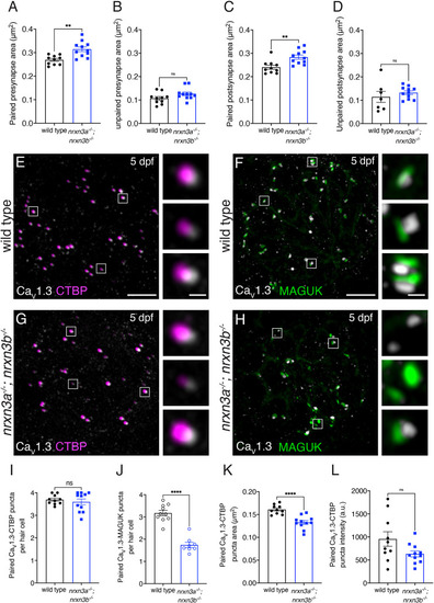
Loss of Nrxn3 impacts pre- and postsynapse size and CaV1.3 channel localization in lateral-line hair cells. (A-D) There is a significant increase in the area of paired (A,C) but not unpaired (B,D) pre- and postsynapses in nrxn3a; nrxn3b mutants compared with wild-type controls. (E-H) Confocal images of mature neuromasts from wild-type controls (E,F) and nrxn3a; nrxn3b mutants (G,H). CTBP labels presynapses with CaV1.3 (E,G) and MAGUK labels postsynapses with CaV1.3 (F,H). The boxed areas (magnified on right) show three examples of individual synapses. Hair cells in A-D were visualized with an Otoferlin or Parvalbumin co-label (not shown). (I-L) Quantification shows no change in the number of CaV1.3-CTBP paired puncta per hair cell in nrxn3a; nrxn3b mutants (I). However, there are significantly fewer CaV1.3-MAGUK paired puncta per hair cell in nrxn3a; nrxn3b mutants (J). The area (K) but not the average intensity (L) of CaV1.3 puncta associated with CTBP puncta is significantly lower in nrxn3a; nrxn3b mutants. n=10 wild-type and 11 nrxn3a; nrxn3b mutant neuromasts in A-D,I,K,L and n=10 wild-type and 8 nrxn3a; nrxn3b mutant neuromasts in J. Images and quantification are from larvae at 5 dpf. An unpaired two-tailed t-test was used in A-D and I-L. ns, P>0.05; **P<0.01; ****P<0.0001. Data are mean±s.e.m. Scale bars: 5 µm (E-H); 1 µm (E-H, insets).

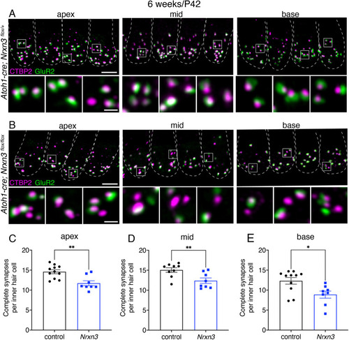
NRXN3 is required at 6 weeks for proper synapse number in mouse auditory inner hair cells. (A,B) Confocal images of mouse inner hair cells (IHCs) at 6 weeks (P42) from control (A) and Nrxn3 mutant animals (Atoh1-Cre; Nrxn3flox/flox) (B). CTBP2 labels the presynapses and GluR2 labels the postsynapses. Merged images show four IHCs from three different regions of the cochlea (apex, middle and basal thirds) for each genotype. Dashed lines outline hair-cell bodies [via a MYO7A co-label (not shown)]. White boxes in the top panels are magnified below to highlight synapses more clearly. (C-E) Quantification reveals that, compared with controls, Nrxn3 mutants have significantly fewer complete synapses per IHC at the apex (C), mid (D) and base (E). These findings were compiled from four animals from each genotype and from two independent litters and immunostains. Each dot represents the average synapse number from one imaging region (6-9 IHCs). Two imaging regions were examined per animal for each tonotopic region. An unpaired two-tailed t-test was used in C-E. *P<0.05; **P<0.01. Data are mean±s.e.m. Scale bar: 5 µm (A,B); 1 µm (A,B, insets).

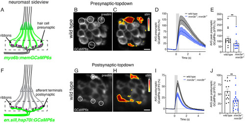
Nrxn3 is required for proper hair-cell synapse function in the lateral line. (A) Side view schematic of a neuromast expressing memGCaMP6s in hair cells. The dashed box indicates the region used to measure presynaptic GCaMP6s responses. (B,C) ΔF heatmaps showing presynaptic GCaMP6s increases in hair cells before (B) and during (C) a 500 ms fluid-jet stimulation in a wild-type neuromast. ROIs (circled) indicate synaptically active hair cells. The black area in the center of each cell is the nucleus. (D) ΔF/F0 GCaMP6s traces showing average presynaptic responses during stimulation for wild-type controls (black) and nrxn3a; nrxn3b mutants (blue). (E) Maximum ΔF/F0 presynaptic calcium responses are significantly reduced in nrxn3a; nrxn3b mutants. n=15 wild-type and 14 nrxn3a; nrxn3b mutant neuromasts at 5-6 dpf. (F) Side view schematic of a neuromast expressing GCaMP6s in afferent terminals beneath hair cells. The dashed box indicates the region used to measure postsynaptic GCaMP6s responses. (G,H) ΔF heatmaps showing postsynaptic GCaMP6s increases in afferent terminals before (G) and during (H) a 500 ms fluid-jet stimulation in a wild-type neuromast. ROIs (circled) indicate synaptically active terminals. (I) ΔF/F0 GCaMP6s traces showing average postsynaptic responses during stimulation for wild-type controls (black) and nrxn3a; nrxn3b mutants (blue). (J) Maximum ΔF/F0 postsynaptic calcium responses to stimulation for wild-type controls and nrxn3a; nrxn3b mutants. n=13 wild-type and 22 nrxn3a; nrxn3b mutant neuromasts at 4-5 dpf. Traces in D and I are displayed as mean, dashed lines are s.e.m.; gray bar denotes the stimulus. Each dot in E and J represents the average response from a single neuromast. A Mann–Whitney U-test was used in E and an unpaired two-tailed t-test was used in J. **P<0.01. Data are mean±s.e.m. Scale bars: 5 µm.

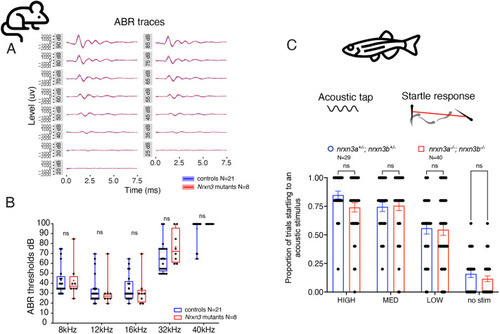
Auditory behaviors are unaltered in mouse and zebrafish after loss of Nrxn3. (A) Average auditory brainstem responses (ABRs) from control and Nrxn3 mutant animals (Atoh1-Cre; Nrxn3flox/flox) at P28-P32 from 20 to 90 dB are shown. (B) No difference was observed between control and Nrxn3 mutants with regards to the ABR threshold at any frequency tested. n=21 control and 8 Nrxn3 mutants. Distributions are framed with 25-75% whisker boxes where exterior lines show the minimum and maximum, the middle line represents the median, and + represents the mean. (C) A vibrational acoustic tap stimulus was used at three stimuli of decreasing intensity to trigger an escape response in nrxn3a+/−; nrxn3b+/− double heterozygotes and nrxn3a−/−; nrxn3b−/− double mutants. The proportion of times (out of five trials) an animal responded to each stimulus is shown. No difference was observed at any stimulus intensity. n=29 nrxn3a; nrxn3b double heterozygotes and 40 nrxn3a; nrxn3b double mutants at 5 dpf. Data are mean±s.e.m. A two-way ANOVA was used in B and C. ns, P>0.05.

Acknowledgments
This image is the copyrighted work of the attributed author or publisher, and ZFIN has permission only to display this image to its users. Additional permissions should be obtained from the applicable author or publisher of the image. Full text @ Development