FIGURE SUMMARY
Title

Morphological, behavioral and cellular analyses revealed different phenotypes in Wolfram syndrome wfs1a and wfs1b zebrafish mutant lines

Authors
Crouzier, L., Richard, E.M., Diez, C., Alzaeem, H., Denus, M., Cubedo, N., Delaunay, T., Glendenning, E., Baxendale, S., Liévens, J.C., Whitfield, T.T., Maurice, T., Delprat, B.
Source
Full text @ Hum. Mol. Genet.

Characterization of wfs1 mutant zebrafish lines. (A) Schematic molecular structure of Wfs1a and Wfs1b proteins with mutation. The stars represent the punctual mutations, blue for Wfs1a mutation and orange for Wfs1b. (B) Relative wfs1a and wfs1b mRNA levels assessed by qPCR in wfs1aC825X and wfs1bW493X zebrafish larvae, normalized against zef1α reference gene. (C) Schematic representation of the different measurements of the larva. (D) Representative images of wild-type and homozygous mutant wfs1 larvae. Images to the right show the ear in the wild-type (upper image) and wfs1aC825X mutant (the lower image). (E) Measurement of body size, (F) eye diameter, (G) ear area in lateral view, (H) anterior and (I) posterior otoliths area. Scale bars, 200 μm in (D), 10 μm in the insets. Error bars represent mean ± SEM and the number of fish is indicated below the columns. **P < 0.01, ***P < 0.0001; unpaired t-test.

Expression of wfs1a and wfs1b mRNA in the zebrafish embryo, larva and juvenile. In situ hybridizations showing the expression patterns of wfs1a (left-hand and middle columns) and wfs1b (right-hand column) between 24 hpf and 10 dpf. All images are lateral views with anterior to the left, except for the inset showing expression of wfs1b in the pancreas (dorsal view, anterior to the left) and the sections through the retina. The position of the transverse hand-cut sections through the trunk at 24 and 96 hpf (dorsal to the top) is marked by a thin line in the preceding image. The eyes have been removed from the 10 dpf embryos, to show expression in the mouth and brain. The hand-cut thick sections through the retina, and the second image of the ear at 10 dpf, were taken from samples stained using an alternative protocol that resulted in stronger staining intensity (see Materials and Methods). Expression of wfs1a in the retina at 7 dpf is strong in the photoreceptors, outer plexiform and inner nuclear layers but is weaker or missing from the inner plexiform and ganglion cell layers (GCLs) and is very weak or absent from displaced amacrine cells (arrowhead). Expression of wfs1b in the retina at 7 dpf appears ubiquitous. Expression in the inner ear appears ubiquitous for both genes and may include some trapping. All images are bright-field images apart from the images of the trunk at 24 hpf, pectoral fin at 48 hpf and retinae at 7 dpf, which were taken with DIC optics. Abbreviations: ac, anterior crista; ascc, anterior semicircular canal; ep, epithelial projections in the otic vesicle; gcl, ganglion cell layer; h, heart; hb, hindbrain; inl, inner nuclear layer; ipl, inner plexiform layer; lc, lateral crista; lev-5, levator arcus branchialis 5; mp, muscle pioneer cells; opl, outer plexiform layer; p, pancreas; pc, posterior crista; pf, pectoral fin; pr, photoreceptors; pscc, posterior semicircular canal; rpe, retinal pigmented epithelium; s, somites; sb, swimbladder; sm, slow-twitch muscle fibers. Scale bars, 200 μm in all images.

Behavioral analyses of 5 dpf wfs1 mutant larvae. (A) Light protocol: the activity is measured for 70 min, with 30 min of training in the dark (OFF), then two cycles of light/dark (ON/OFF) of 10 min each. Analysis of the distance traveled by (B) the wfs1aC825X and (F) wfs1bW493X larvae at 5 dpf during the light/dark cycle in the VMR test. Relative distance traveled was measured during some experiment phases as (C, G) the training phase [blue-dotted lines in (B, F) between 21 and 29 min]; (D, H) the ON phase (the averaged ON1 and ON2 phases); (E, I) the OFF phase (the averaged OFF1 and OFF2 phases). (J) Illustration of the OKR test. Four larvae are immobilized in petri dish and placed in an arena with moving black and white gratings. (K) The number of saccades performed in 2 min. (L) Illustration of the touch escape test. The larva’s tail is touched with a tip and (M) the traveled distance of the larva in the rail is measured for 5 s, repeated for three times per larva and averaged. Relative distances were expressed as % of associated controls. Error bars represent ± SEM calculated from three replicas. The number of animals is indicated below the columns. *P < 0.05; **P < 0.01; unpaired t-test.

Analysis of the quantity of movement of 5 dpf wfs1 mutant larvae during the noise cycle in the ASR test. (A) Sound protocol: the activity is measured for 45 min in a total dark (OFF) condition, with a training phase for 30 min silently, then three cycles of white sounds (90 dB) of 1 s each and 5 min interspersed. Quantity of movement per second for (B) the wfs1aC825X and (F) wfs1bW493X larvae according to the sound protocol. Relative quantity of movement during (C, G) the training phase [blue-dotted lines in (B, F) between 21 and 29 min]; (D, H) the baseline phase, period of 2 min before each sound (the averaged three baseline phases); (E, I) the three stimulations (the averaged sounds phases). Activity was expressed as % of associated controls. Error bars represent ± SEM calculated from three replicas. The number of animals is indicated below the columns. *P < 0.05; unpaired t-test.

Molecular characterization of the retina and measure of the motor neuron length for both wfs1 mutant zebrafish lines. Retina measurements in wfs1 mutants with (A) quantification of the number of ganglion cells, (B) the thickness of the associated layer and (C) photoreceptors cells as red and green cones labeled with Zpr-1 antibody and (D) rods with anti-Rho4d2 antibody. Quantification of the tail motor neuron length of 24 hpf (E) and 48 hpf (F) zebrafish. For each fish, five to six neurons were measured, and the average length was calculated. Scale bar, 50 μm. The number of animals is indicated below the columns. *P < 0.05; ***P < 0.001; unpaired t-test.

Relative mRNA and protein expression levels of larvae zebrafish ER stress factors in physiological condition at 5 dpf. mRNA levels were analyzed by qPCR and protein contents by western blot in (A, C) wfs1aC825X and (B, D) wfs1bW493X. zeif2α and Stain-free™ were used as controls in qPCR and western blot analysis, respectively. The expression levels of mRNA and protein are represented as the fold change from the control group. Data are expressed in mean ± SEM, n = 5–9 in each group. *P < 0.05, ***P < 0.001; unpaired t-test.

Relative gene expression levels of ER stress factors in zebrafish larvae at 5 dpf exposed to tunicamycin during 24 h. Expression analysis of the selected genes using cDNA prepared from wfs1aC825X (A) and wfs1bW493X (B) zebrafish larvae. The relative expression levels of mRNA are represented as the fold change from the control vehicle group. Data are expressed in mean ± SEM, n = 5 in each group. *P < 0.05, **P < 0.01, ***P < 0.001 versus DMSO condition, °P < 0.05, °°°P < 0.001 versus WT condition; two-way ANOVA, unpaired t-test.

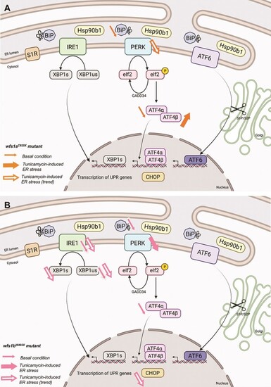
Schematic summary of ER stress pathways alterations observed in basal condition or after tunicamycin treatment in wfs1aC825X (A) and wfs1bW493X (B) zebrafish line.

Analysis of mitochondrial respiration in zebrafish larvae at 2 and 5 dpf with Seahorse XF mito stress test. OCR profiles of (AL) wfs1aC825X at 2 dpf (A–F) and 5 dpf (G–L); and OCR profiles of (MX) wfs1bW493X at 2 dpf (M–R) and 5 dpf (S–X) during the assay, with (B, H) basal respiration, (C, I) ATP production, (D, J) maximal respiration, (E, K) proton leak and (F, L) non-mitochondrial respiration. Error bars represent ± SEM calculated from three replicas. The number of animals is indicated below the columns. *P < 0.05, **P < 0.01; unpaired t-test.

Acknowledgments
This image is the copyrighted work of the attributed author or publisher, and ZFIN has permission only to display this image to its users. Additional permissions should be obtained from the applicable author or publisher of the image. Full text @ Hum. Mol. Genet.