FIGURE SUMMARY
Title

Loss of rps9 in Zebrafish Leads to p53-Dependent Anemia

Authors
Chen, C., Huang, H., Yan, R., Lin, S., Qin, W.
Source
Full text @ G3 (Bethesda)

ZFIN is incorporating published figure images and captions as part of an ongoing project. Figures from some publications have not yet been curated, or are not available for display because of copyright restrictions.

Embryos with rps9 deficiency display morphological abnormalities. (A-E) Overall morphological phenotype of rps9 deficient embryos. At 24 hpf (A and A’), smaller eyes and abnormal hindbrain (arrowhead) appear in the head region. By 36 hpf (B), enlarged hindbrain (arrowhead) becomes apparent and pigment is reduced. From 48 hpf to 4 dpf (C-E), edema (arrowhead) is gradually developed in the pericardial region, accompanied with shortened trunk. (F) Cartilage fails to grow in the pharyngeal arch in rps9 mutant embryos.

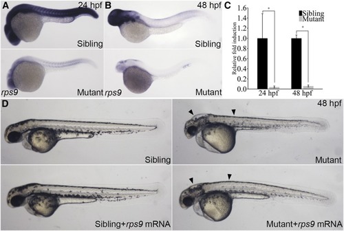
Phenotype of rps9 deficient embryos is partially rescued by rps9 mRNA injections. (A-B) Whole mount in situ hybridization for rps9 in mutants and siblings at 24 hpf or 48 hpf. (C) qPCR analysis of rps9 transcript levels. *, P < 0.05. (C) In vitro transcribed rps9 mRNA (400 pg) partially rescues phenotype in rps9 mutants. Arrowhead indicates morphological improvement in the hindbrain and trunk.

Deficiency in rps9 induces anemia in mutant embryos. (A-B) Detection of hemoglobin levels by o-dianisidine staining for rps9 in both siblings and mutants at 48 hpf and 3 dpf. (C-E, E’) Detection of erythroid cells by crossing rps9 mutant into LCR:EGFP transgenic fish line. At 24 hpf, the number of blood cells are reduced slightly; At 48 hpf and 3 dpf, erythrocytes are almost deprived in the trunk, and only a small amount of red blood cells remain in the cardiac region. (F) Wright-Giemsa staining of erythroid cells at 3 dpf in sibling and mutant.

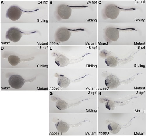
No obvious change was seen for expression of erythroid specific markers in rps9 mutants. (A-F) Expression levels of gata1, hbbe1.1, and hbae3 in mutants remain unchanged at 24 hpf (A-C) and 48 hpf (D-F). (G-H) Because of the shortened size of the mutant embryos, slight decrease in hbbe1.1 and hbae3 is seen at 3 dpf.

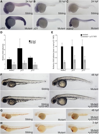
Anemic phenotype is partially rescued by p53 konckdown in rps9 deficient embryos. (A-C) Whole mount in situ shows that p53, p21, and mdm2 are upregulated in mutants. (D) The expression of p53 and p53 target genes including mdm2 and p21 increases as measured by qPCR. The relative fold induction is represented as fold change over sibling control. (E) p53 inhibition by MO injection knockdown the gene expression of p53 and p53-target genes. (F) The morphological defects of rps9 mutant are largely rescued by p53 inhibition. (G-H) An increase of hemoglobin level appears at 48 hpf. (G) and 3 dpf (H) after p53 knockdown.

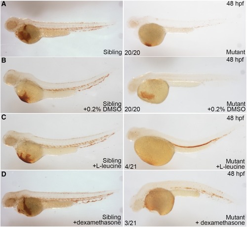
Hemoglobin level is partially recovered by L-leucine, and dexamethasone treatment in rps9 deficient embryos. (A-B) Mutants without treatment and mutants treated with 0.2% DMSO were used as controls. (C) Mutants treated with L-leucine (200 mmol/L) show a considerable recovery in hemoglobin level. (D) Mutants treated with dexamethasone (250 μmol/L) display a less effective restoration in hemoglobin level.

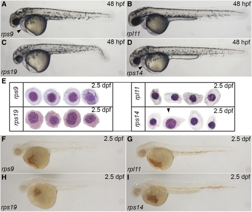
The severity of phenotype of four ribosomal protein mutants increases in an order of rpl11, rps14, rps9, and rps19. (A-D) Morphological comparison of four mutants by 48 hpf. Arrowhead indicates pericadial anema. (E) Wright-Giemsa staining for four mutants at 2.5 dpf. Arrowhead indicates that an erythrocyte in rps14 mutants resembles the erythrocytes in rps9 mutants. (F-I) o-dianisidine staining of four mutants at 2.5 dpf.

Unillustrated author statements

PHENOTYPE:
Fish:
Observed In:
Stage Range: Prim-5 to Day 4
Acknowledgments
This image is the copyrighted work of the attributed author or publisher, and ZFIN has permission only to display this image to its users. Additional permissions should be obtained from the applicable author or publisher of the image. Full text @ G3 (Bethesda)