- Title
-
Neurodegeneration and Epilepsy in a Zebrafish Model of CLN3 Disease (Batten Disease)
- Authors
- Wager, K., Zdebik, A.A., Fu, S., Cooper, J.D., Harvey, R.J., Russell, C.
- Source
- Full text @ PLoS One
|
cln3 is expressed in the developing WT zebrafish. A-N) In situ hybridisation for cln3 (antisense) shows its expression from the one cell stage through to 4 dpf. From 1 dpf expression is increasingly restricted to the central nervous system. At 2 dpf cln3 expression is particularly strong in the mid-hindbrain boundary (arrow) where the cerebellum is developing (G, H, M, N). (A'-L') Experiments conducted using a cln3 sense probe (sense) revealed the specificity of the antisense signal. Abbreviations: mhb, mid-hindbrain boundary. |
|
cln3 ATG MO morphants have abnormal brain and heart morphology. (A, A') 32 hpf normal development of the WT forebrain, midbrain, hindbrain, retina and fourth ventricle. (B, B') 32 hpf 1.6 ng cln3 ATG MO morphants display abnormal development of all parts of the brain, a smaller retina, and enlargement of the fourth ventricle. (C, C') normal development of the WT tail, yolk, pericardial sac and heart at 4 dpf. The WT brain completely fills the cranium. (D, D') 1.6 ng cln3 ATG MO morphants have a curved tail, and a larger yolk and pericardial sac at 4 dpf. The heart has an elongated appearance and is lacking pigmented erythrocytes. The fourth ventricle is enlarged and the mid- and hindbrain appear smaller. Lateral views. Anterior is to left. Dorsal is up. Abbreviations: v, fourth ventricle; hb, hindbrain; mhb, mid-hindbrain boundary; mb, midbrain, fb, forebrain, r, retina; t, tail; y, yolk; ps, pericardial sac; h, heart; (n = 4 per group). Lateral views. Scale bars: A-D 250 ?m; A'-D' 100 ?m. |

ZFIN is incorporating published figure images and captions as part of an ongoing project. Figures from some publications have not yet been curated, or are not available for display because of copyright restrictions. PHENOTYPE:
|

ZFIN is incorporating published figure images and captions as part of an ongoing project. Figures from some publications have not yet been curated, or are not available for display because of copyright restrictions. |
|
Neurons and glia are disrupted in cln3 ATG MO morphants. (A-A'', B-B'') Immunohistochemical staining for axons (acetylated ?-tubulin) at 4 dpf. (A-A'') normal development of axons in WT larvae. (B-B'') 1.6 ng cln3 ATG MO morphants have a complete absence of axonal organisation throughout the brain, with axonal accumulation (B, dashed arrows), loss of the optic tectum and a narrowing of the optic nerve (B'', dashed arrow). (A-A'', B-B''), anterior to the left; A, B, lateral view, dorsal up; A', B', dorsal view; A'', B'' ventral view. (C-C', D-D') Immunohistochemistry using antibodies to glia (glial fibrillary acidic protein, GFAP) at 4 dpf. (C-C'') Normal staining in WT larvae. (D-D') Ectopic GFAP is observed in the notochord in 1.6 ng cln3 ATG MO morphants (dashed arrow). Lateral view. Anterior to the left. Dorsal up. (E-E', F-F', G-G') Transgenic zebrafish expressing GFP under the control of the HuC promoter in neurons were injected with 1.6 ng cln3 ATG MO and observed at 3 dpf. (E-E') In WT zebrafish, the normal structure of the developing brain and retina can be observed. (F-F') In morphants, there appear to be fewer neurons and the normal brain structure is lost. Many GFP-positive cells were enlarged and found nearer the surface of the brain (F', dashed arrows). (G-G') When the morphology of these enlarged cells was examined further, they lacked typical neuronal morphology. Lateral view. Anterior to the left. Dorsal up. Abbreviations: cb, cerebellum, fb, forebrain; hb, hindbrain; ot, optic tectum; on, optic nerve. A-G'' (all images) Z projection. Scale bars: 100 ?m. n = 4 per group. PHENOTYPE:
|
|
Cellular proliferation is abnormal in cln3 ATG MO morphant zebrafish. (A, A') Proliferation, assayed at 4 dpf using anti-PH3 (a marker of proliferative cells in mitotic M phase), is observed throughout the 4 dpf WT retina, jaw and the brain. (B, B') A marked reduction in the amount of cellular proliferation throughout the retina can be seen in the 1.6 ng cln3 ATG MO morphant. Although not quantified, it appears that proliferation in the morphant brain (B) is increased compared to WT. Confocal images are Z-projections. Scale bar: 100 ?m (A, A', B) and 50 ?m (B'). Lateral views. Anterior is to the left. Dorsal is up. (C) Quantification of these data show that the number of proliferating cells in the morphant retina is significantly reduced from 100.3 cells in WT to 50.3 cells in morphants; ***p<0.0006 (n = 3 zebrafish per group). (D) Quantification demonstrating a significantly reduced mean retinal area in the morphants (0.0566 mm2 for WT retinae compared to 0.0135 mm2 for morphant retinae; ****p<0.0001 (n = 10 zebrafish per group)). (C, D) Data represent mean ąSD; results were evaluated using a 2-tailed unpaired Student's t-test. PHENOTYPE:
|
|
Apoptotic cells and lysosomal storage were found in the cln3 ATG MO morphant brain. Acridine orange and Lysotracker staining was carried out on live WT and cln3 ATG MO morphant zebrafish aged 4 dpf. (A-A'') WT fish showed low levels of programmed cell death in the brain and very little bright lysosomal staining. (B-B'') Morphants injected with 1.6 ng of cln3 ATG MO had increased levels of programmed cell death (acridine orange, bright green) in the brain, this localised particularly to the optic tectum, but only a slight increase in lysosomal staining (red). (C-C'') Morphants injected with 2.9 ng of cln3 ATG MO showed very high levels of programmed cell death in the forebrain, midbrain, retina and yolk-body boundary; lysosomal hypertrophy was also observed. (D-D'') Morphants injected with 2.9 ng of cln3 ATG MO exhibit abundant apoptotic bodies in the forebrain that often co-localises with lysosomes (arrows). Bright green, acridine orange in apoptotic bodies; Red, Lysotracker red in lysosomes. Abbreviations: ab, apoptotic body; lys, lysosome; ot, optic tectum. A-A", B-B", C-C'' are Z projections. D-D'' are the same Z slice. Lateral views. Anterior to the left. Dorsal up. The scale bars represent 200 ?m and apply to all panels. PHENOTYPE:
|
|
Subunit c accumulates in lysosomes, and mitochondria are compromised in cln3 ATG MO morphants. (A-A", B-B") Immunohistochemical staining for lysosomal associated membrane protein 1 (LAMP1, green) and subunit c of the mitochondrial ATP synthase (subunit c, red) at 4 dpf. (A-A'') WT. (B-B'') 1.6 ng cln3 ATG MO morphants. In morphants, lysosomes appear larger (B, dashed arrow), subunit c accumulates (B', dashed arrow) and they co-localise (B'', dashed arrows). Abbreviations: nc, notochord. Z slice. Lateral view. Anterior to left. Dorsal up. Scale bars: 50 ?m. (C-C', D-D') Mitotracker stain labelling mitochondria in superficial cells of the eye at 4 dpf. (C-C') WT. (D-D') 1.6 ng cln3 ATG MO morphant. In WT cells, many individual mitochondria are observed, whereas in morphant cells the stain has not been accumulated by mitochondria suggesting loss of mitochondrial membrane potential. Z slice. Lateral view. Scale bars: C, D 25 ?m; C', D' 10 ?m. PHENOTYPE:
|
|
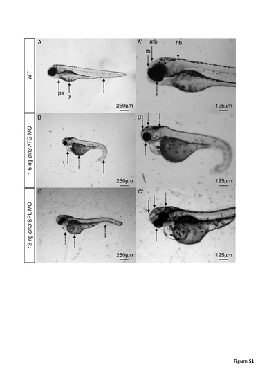
Abnormal morphology was observed using two different cln3 morpholinos at 4 dpf. (A, A') WT. (B, B') 1.6 ng cln3 ATG MO. (C, C') 12 ng cln3 SPL MO. Morphant larvae (B, B', C, C') showed small retinas, small brain, pericardial oedema, a large yolk sac and abnormal tail curvature (dashed arrows) compared to WT (A, A'). Abbreviations: r, retina; ps, pericardial sac; y, yolk; t, tail; fb, forebrain; mb, midbrain; hb, hindbrain. Lateral views. Anterior to left. Dorsal up. Scale bars: A-C 250 ?m; A'-C' 125 ?m. PHENOTYPE:
|

