- Title
-
Aromatic L-Amino Acid Decarboxylase (AADC) Is Crucial for Brain Development and Motor Functions
- Authors
- Shih, D.F., Hsiao, C.D., Min, M.Y., Lai, W.S., Yang, C.W., Lee, W.T., and Lee, S.J.
- Source
- Full text @ PLoS One
|
ddc expression pattern in zebrafish. ddc mRNA was expressed from ubiquitous to specific areas in brain during whole developmental stage. Whole mount RNA in situ hybridization was performed using a ddc-specific anti-sense probe on embryos at (A?B) shield stage, (C?D) bud stage, (E?F) 14 hpf, (G?H) 18 hpf, (I?J) 24 hpf, (K?L) 36 hpf, (M?N) 48 hpf, and (O?P) 74 hpf. (Q) was amplified from 36 hpf, 48 hpf, and 74 hpf, respectively. Panels (A), (C), (E), (G), (I), (K), (M) and (O) are dorsal view. The rest are lateral view. Arrowheads, epiphysis (Epi); arrows, direct locus coeruleus (LC); brackets, diencephalic catecholaminergic cluster (DCA); staining in square, raphe nuclei (RN). (R?S) The RT-PCR analysis for ddc expression in time scan and Adult tissue scan. |
|
Inhibition of AADC by NSD-1015 in zebrafish. AADC inhibitor NSD-1015 caused morphological and behavioral defects. Compared to untreated fish, the inhibited larvae became shorter by gradient increase of NSD-1015 concentration. (A) The phenotype of treated larvae. From up to bottom is the untreated larvae (n=234), and treated larvae by inhibitor NSD-1015 100 μM(n= 312), 200 μM (n=300), 300 μM (n=304), 400 μM (n=264), and 500 μM (n=260). (B?C) The statistics of body length and movement in touch response, respectively, and showed the decrease of body length and movement in touch response in larvae treated with NSD-1015. (D) Sequencing touch response for untreated (upper panel) and treated larvae (bottom panel), showing decreased movement in touch response in larvae treated with NSD-1015. Results were expressed as meanąSD. **p<0.01, ***p<0.001 compared to control larvae. |
|
Morphologic and behavioral changes observed in ddc morphants. The change of body length (A) and touch response (B) in control (n=124) and 0 (n=123), 1.25 ng (n=104), 2.5 ng (n=93), 5 ng (n=111) and 10 ng (n=96) tMO1-treated larvae, showing the dose-dependent decrease of body length and touch response in treated larvae. (C) Brain volume calculation showing the decrease of brain volume compared with control, in 1.25, 2.5, 5 and 10 ng tMO1-treated larvae (n=7 in each test). (D) Compared with control and buffer image stained with Toluidine blue, horizontal section in 3 dpf morphants revealed smaller brain size in 2.5, 5 and 10 ng morphants. DT, dosal thalamus; poc, postoptic commissure; PT, posterior tuberculum; TeO, tectum opticum. Statistical analyses of brain to white matter ratio (E) and brain cell numbers (F) in control, and 0, 1.25, 2.5, 5 and 10 (n=7 each) ng tMO1-treated larvae are shown. The brain area is enclosed by a dash line boundary and the white mater area is light blue staining areas within the brain. All results were expressed as meanąSD. *p<0.05, **p<0.01, ***p<0.001 compared to control larvae. PHENOTYPE:
|
|
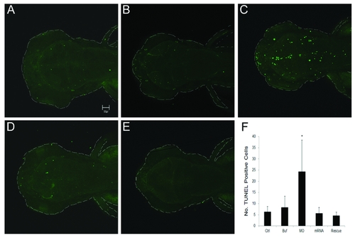
TUNEL staining for apoptosis in treated larvae. All larvae were evaluated at 3 dpf. (A) Control. (B) Injection of 1x Danieaus? buffer with 1% phenol red. (C) Injection of 5 ng ddc tMO1. (D) Injection of 100 pg ddc mRNA. (E) Injection of mixture of 5 ng ddc tMO1 and 50 pg ddc mRNA with non-MO1 binding site. Apoptotic cells were increased in ddc morphants compared to other controls. The increase of apoptosis can be attenuated by co-injection of ddc mRNA. (F) Statistical analysis. Photographs were taken by Zeiss Confocal 700 microscope at 100X magnification. All data represented at least three independent experiments and were expressed as meanąSD. *p<0.05 compared to control larvae. PHENOTYPE:
|
|
Morphological changes in DA neuron patterning. In situ hybridization with tyrosine hydroxylase anti-sense mRNA probe. (A) Control. (B) 5 ng ddc tMO1 treated. (C) 100 pg ddc mRNA treated. (D) Co-injected mixture with 5 ng ddc tMO1 and 50 pg ddc mRNA with non-tMO1 binding site. Numbers show the site of dopaminergic neuron clusters 1-7 and 8, which indicated spots in retina, locus coeruleus (LC), pre-tectum (PrC, arrowhead) and olfactory bulb (OB, arrow). Compared to tyrosine hydroxylase expression pattern in control larvae, several tissue parties including olfactory bulb, pre-tectum and DA cluster were malpositioned or absent in the ddc morphant brains. EXPRESSION / LABELING:
PHENOTYPE:
|
|
Behavioral evaluations in 5 dpf morphants showing the reduction of swimming ability. Both total distance and average velocity in swimming behavior were decreased in ddc morphants. (A) Swimming routes of a 5 dpf larva with injection of none, 1x injection buffer, 5 ng tMO1,100 pg mRNA and 5ng tMO1 and 100 pg ddc mRNA mixture to rescue, respectively. The video was recorded for 25 min. (B) Statistical analysis showed total distance of trajectories. The ddc morphants revealed decreased total distance of swimming compared with different controls. However, co-injection of 100 pg full length ddc mRNA can significantly attenuated the reduction of total swimming distance. (C) Statistical analysis for swimming velocity among different controls and ddc morphants, and rescued larvae also revealed the same results. The bars represent changes in different treatment of 5 dpf larvae in three independent experiments. n=31 for each treated larvae. Results were expressed as meanąSD. *p<0.05 compared to control larvae. PHENOTYPE:
|
|
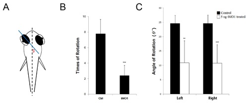
Eye movement in 5 dpf morphant significant reduced during rotation. Both frequency and angle of rotation decline compared with control and morpahant. (A) Schematic representation of the method used to obtain eye movement recordings from embedded larvae with 3% methylcellulose. Calculate with the angle ¸ to plane of eyeball and body axis. (B) Statistics analysis for eye rotation frequency during 120 seconds. (C) Statistics anaylsis for average of eye movement angle. Left: left eyeball, Right: right eyeball. The bars represent changes in different treatment of 5 dpf larvae in three independent experiments. n=7 for each treated larvae. Results were expressed as meanąSD. **p<0.01, ***p<0.001 compared to control larvae. PHENOTYPE:
|
|
Efficiency testing of ddc translational blocking MOs. (A) A 303 bp partial sequence of ddc containing tMO1 or tMO2 targeting site (purple region) was inserted into a pCS2+ vector containing CMV promoter (brown region) and GFP (green region) sequences. The binding sequences for ddc tMO1 and tMO2 are shown below the construct map. (B) Embryos were co-injected with designated MO (5 ng) and MO efficiency testing plasmid (100 pg), examined by epifluorescent microscope under bright and dark fields. Representative superimposed images are shown. (C) Percentages of embryos showing GFP in different treatments are shown. Plasmid injected only group, n=106; tMO1,n=103; tMO2, N=64. * p <0.001. |
|
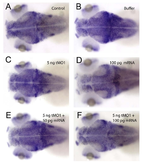
ddc MO caused reduction in brain size as examined by whole-mount in situ hybridization against huc. Embryos were treated as indicated and subjected to whole-mount is situ hybridization against huc. |


