- Title
-
Haploinsufficiency of tumor suppressor pten predisposes to hemangiosarcoma in zebrafish
- Authors
- Choorapoikayil, S., Kuiper, R.V., de Bruin, A., and den Hertog, J.
- Source
- Full text @ Dis. Model. Mech.
|
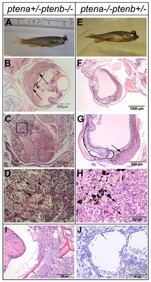
Ocular tumor of ptena +/-ptenb -/- and ptena -/-ptenb+/- mutants, diagnosed as hemangiosarcoma. (A-D) 3-month-old ptena+/-ptenb -/- and (E-H) 9-month-old ptena -/-ptenb+/- mutant with ocular tumor. The entire intact fish was fixed and embedded in paraffin. (B-D) Transversal sections and (F-H) sagittal sections were stained with H&E. Arrows indicate tumor mass, which is associated with the eye bulbs. (B,G) Higher-power magnifications of the tumor mass; (D) magnification of the boxed area in C. The tumor consists of cells that form different sizes of blood-filled spaces (arrows in D,H).(I,J) H&E staining of sections from two individuals revealed hemangiosarcoma formation. (I) The tumor was invasive and penetrated into the brain region with enclosing scull elements (arrows). (J) Cells with plump morphology (arrow) are detaching from surrounding tissue and protrude into the vessel lumen. Sections of representative tumors are depicted here. PHENOTYPE:
|
|
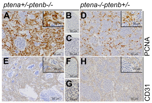
Low-dose Pten tumors display elevated cell proliferation and have endothelial cell origin. ptena+/-ptenb-/- (n=6) (A-C,E-G) and ptena-/-ptenb+/- (n=1) (D,H) mutants with tumors were fixed, paraffin embedded and sectioned transversally or sagitally. Immunohistochemistry using PCNA, a cell proliferation marker, showed clearly enhanced nuclear PCNA staining in tumor cells (A,D) compared with control tissue in the same sections (B,C). CD31, an endothelial cell marker, was expressed in the tumor tissue (E,H) in a similar manner as in control blood vessels in the same sections (F,G). Representative sections are depicted here. EXPRESSION / LABELING:
|
|
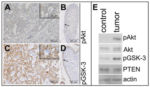
Elevated Akt/PKB signaling in Pten mutant tumors. Immunohistochemistry on transversal sections from a ptena+/-ptenb-/- mutant fish, using pAkt- and pGSK-3β-specific antibodies. (A) Tumor area stains weakly positive for pAkt, whereas (B) control vessel (arrow) from the same section is not stained (n=10). (C) Tumor area is highly positive for pGSK-3β, whereas (D) cells from a control vessel (arrow) from the same section only stain mildly positive (n=6). (E) The cranial part of a ptena+/-ptenb-/- mutant that developed a tumor was dissected into two fragments, one harboring tumor tissue (tumor) and the other representing control tissue (control). The samples were lysed and the lysates were run on a denaturing SDS-polyacrylamide gel. The proteins were transferred to a PVDF membrane and after blocking the blot was probed with anti-pAkt antibody, stripped and sequentially probed with anti-Akt, anti-pGSK-3, anti-PTEN and, as a loading control, anti-actin. EXPRESSION / LABELING:
|
|
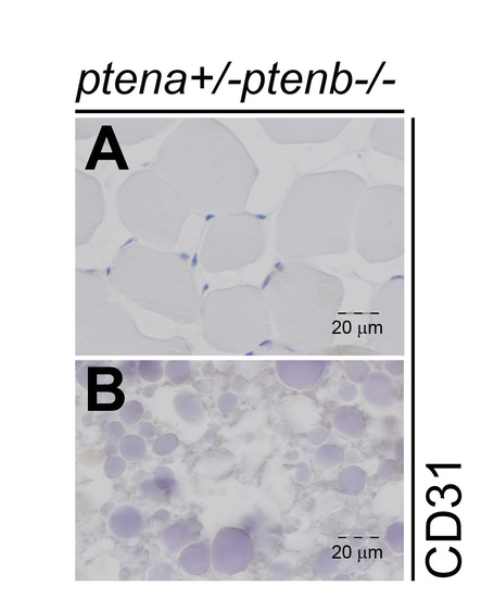
Muscle and ovary tissue do not exhibit CD31 staining. Immunohistochemistry on transversal sections from a ptena+/-ptenb-/- mutant fish with a diagnosed hemangiosarcoma, using CD31 specific antibody. Different areas from the CD31-stained sections in Fig. 4 are depicted here, which contain (a) muscle and (b) ovary. Both do not show CD31 staining, as expected for these non-endothelial tissues.. Sections of representative tissues are depicted. Scale bar is 20 μm, as indicated. |
|
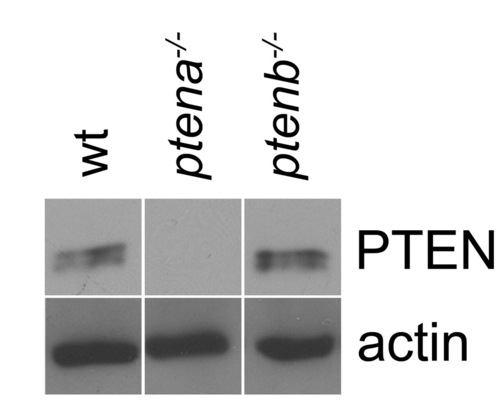
PTEN antibody specifically recognizes Ptena and not Ptenb protein. Protein lysate from 3 dpf wild type, ptena-/- and ptenb-/- embryos were run on an SDS-polyacrylamide gel. The proteins were transferred to a PVDF membrane and after blocking the blot was probed with anti-PTEN antibody, stripped and sequentially probed with actin as a loading control. Detection was by enhanced chemiluminescence and representative blots are shown. Specific signal is detected in wild type and ptenb-/- embryos, but not in ptena-/- embryos, indicating that the PTEN antibody is specific for Ptena. |

