
|
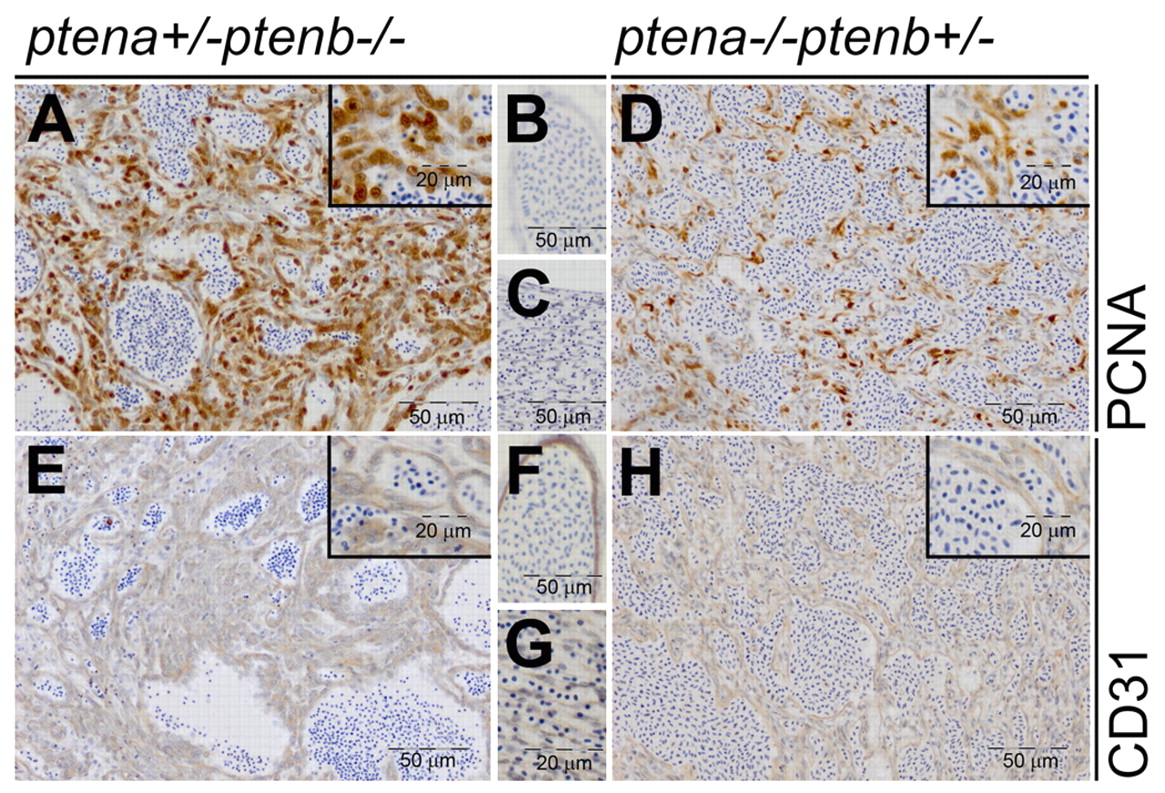
Fig. 3
Low-dose Pten tumors display elevated cell proliferation and have endothelial cell origin. ptena+/-ptenb-/- (n=6) (A-C,E-G) and ptena-/-ptenb+/- (n=1) (D,H) mutants with tumors were fixed, paraffin embedded and sectioned transversally or sagitally. Immunohistochemistry using PCNA, a cell proliferation marker, showed clearly enhanced nuclear PCNA staining in tumor cells (A,D) compared with control tissue in the same sections (B,C). CD31, an endothelial cell marker, was expressed in the tumor tissue (E,H) in a similar manner as in control blood vessels in the same sections (F,G). Representative sections are depicted here.