- Title
-
A Key Role for Poly(ADP-Ribose) Polymerase 3 in Ectodermal Specification and Neural Crest Development
- Authors
- Rouleau, M., Saxena, V., Rodrigue, A., Paquet, E.R., Gagnon, A., Hendzel, M.J., Masson, J.Y., Ekker, M., and Poirier, G.G.
- Source
- Full text @ PLoS One
|
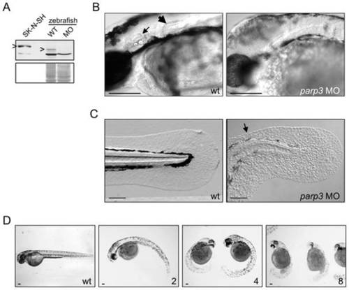
Developmental perturbations in zebrafish embryos with impaired parp3 expression. A. Immunoblot analysis (upper panel) of zebrafish Parp3 in wild type (WT) and parp3 morphants (MO) using an antibody raised against human PARP3. A whole cell extract of human SK-N-SH cells is shown as a control. The protein bands corresponding to PARP3 are indicated by ?>?. The faster migrating band corresponds to a non-related protein that cross-reacts with the antibody. The Western blot membrane was stained with Ponceau S as a protein loading control (lower panel). B. Enlarged lateral views of the head regions of wild type and parp3 morphants injected with 4 ng MO1. The inner ears (small arrow) and the pectoral fins (large arrow) in the wt embryos are not formed in the parp3 morphants. C. Enlarged lateral views of the tail of wild type and parp3 morphants injected with 4 ng MO1. The median fin fold (arrow) is less developed in the morphants and has a more granular aspect. Effects are more pronounced on the dorsal side (arrow). D. Zebrafish embryos 48hrs after injection of increasing amounts of the parp3-specific morpholino oligonucleotide MO1 at the one-cell stage (ng amounts indicated in the lower right corner). The short length of morphant embryos, their curved tail and their reduced pigmentation is increasingly severe with increasing amounts of injected MO1. Lateral views with anterior to the right and dorsal to the top. Scale bars represent 10 μm. |
|
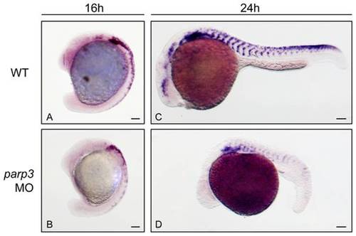
Expression of the neural crest cell marker crestin is impaired in parp3 morphants. Zebrafish embryos were untreated (WT) or injected with 4 ng parp3 MO1 and crestin expression was monitored by in situ hybridization. A. In 16 hpf WT embryos, crestin is expressed in premigratory neural crest cells and in neural crest cells migrating in the anterior trunk segments. B. In 16 hpf parp3 morphants, crestin expression appears to be generally reduced. C. By 24 hpf, crestin-positive cells are distributed along the anterior-posterior axis, in neural crest migratory pathways of WT embryos. D. In 24 hpf parp3 morphants, crestin expression is no longer detectable in the head and is markedly reduced in the trunk. Lateral views of embryos are shown with anterior to the left and dorsal to the top. Scale bars represent 10 μm. EXPRESSION / LABELING:
PHENOTYPE:
|
|
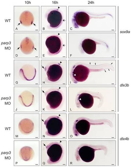
Impaired expression of sox9a, dlx3b and dlx4b in parp3 morphants. Zebrafish embryos were untreated (WT) or were injected with 4 ng parp3 MO1. Gene expression was detected by in situ hybridization. A?F. The expression of sox9a is drastically reduced in the otic placodes (small arrows) at 10 hpf and in the otic vesicles (arrowheads) at 16 hpf. Expression of sox9a in somite cells (small arrows in B and E) appears diffuse in parp3 morphants. Expression of sox9a is almost completely abolished in the head region at 24 hpf (C, F). Expressions of dlx3b (G?L) and dlx4b (M?R) are minimally affected by parp3 MO in ectodermal cells at 10 hpf (G, J, M, P) but are significantly reduced in the otic vesicles (arrowheads), olfactory placodes (large arrows) and branchial arches (white arrows) of parp3 morphants at 16 hpf (H, K, N, Q) and 24 hpf (I, L, O, R). The expression of dlx3b and dlx4b is abolished in the median fin fold of 24 hpf parp3 morphant embryos (small arrows in I). Dorsal views of embryos with anterior to the bottom in A, D, G, J, M, P and lateral views with anterior to the left, dorsal to the top, in B, C, E, F, H, I, K, L, N, O, Q and R. Scale bars represent 10 μm. |
|
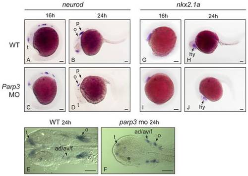
Expression of neurod and nkx2.1 in parp3 morphants. Expression of: A?F, neurod and G?J, nkx2.1a were determined in uninjected embryos or in embryos that received 4 ng of parp3 MO1, by in situ hybridization. A?D and G?J are lateral views with anterior to the left, dorsal to the top. E?F are dorsal views of flat-mounted embryos with anterior to the left. ad/av/f: anterodorsal/anteroventral lateral line/facial placodes/ganglia; p: posterior lateral line placode; e: eye; o: octavel/statosacoustic ganglia precursors; t: telencephalon. Scale bars represent 10 μm. EXPRESSION / LABELING:
PHENOTYPE:
|

Unillustrated author statements |


