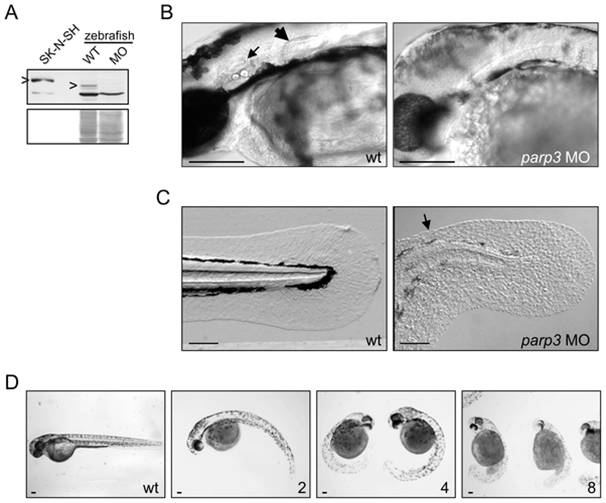
Fig. 1 Developmental perturbations in zebrafish embryos with impaired parp3 expression.
A. Immunoblot analysis (upper panel) of zebrafish Parp3 in wild type (WT) and parp3 morphants (MO) using an antibody raised against human PARP3. A whole cell extract of human SK-N-SH cells is shown as a control. The protein bands corresponding to PARP3 are indicated by “>”. The faster migrating band corresponds to a non-related protein that cross-reacts with the antibody. The Western blot membrane was stained with Ponceau S as a protein loading control (lower panel). B. Enlarged lateral views of the head regions of wild type and parp3 morphants injected with 4 ng MO1. The inner ears (small arrow) and the pectoral fins (large arrow) in the wt embryos are not formed in the parp3 morphants. C. Enlarged lateral views of the tail of wild type and parp3 morphants injected with 4 ng MO1. The median fin fold (arrow) is less developed in the morphants and has a more granular aspect. Effects are more pronounced on the dorsal side (arrow). D. Zebrafish embryos 48hrs after injection of increasing amounts of the parp3-specific morpholino oligonucleotide MO1 at the one-cell stage (ng amounts indicated in the lower right corner). The short length of morphant embryos, their curved tail and their reduced pigmentation is increasingly severe with increasing amounts of injected MO1. Lateral views with anterior to the right and dorsal to the top. Scale bars represent 10 μm.

