- Title
-
Smad2/3 activities are required for induction and patterning of the neuroectoderm in zebrafish
- Authors
- Jia, S., Wu, D., Xing, C., and Meng, A.
- Source
- Full text @ Dev. Biol.
|
Effects of dnsmad3b overexpression on the expression of early neural and non-neural markers. (A?L) Embryos were injected at the one-cell stage at indicated doses and examined by in situ hybridization for marker gene expression at around 60% epiboly stage. Embryos were positioned laterally with dorsal to the right and animal pole to the top. Note that dnsmad3b injection resulted in reduction of the neural markers sox2 and sox3, but increase of the ventral marker gata2. (M?O) Bar graphs showing percentage of embryos with different expression levels of the markers. Normal, decreased or increased expression was indicated by blue, yellow or red colors. The number of observed embryos was indicated in the parentheses. (P?R) Quantitative RT-PCR results of sox2 (P), sox3 (Q) and gata2 (R). The fold change in transcript levels (Y-axis) was graphed relative to control embryos and the data represent the mean ± s.d. from triplicates. The symbols ∗ or ? represent statistical significance at p ≤ 0.05 or insignificance at p > 0.05 compared to the uninjected control or linked samples, respectively. EXPRESSION / LABELING:
|
|
dnsmad3b interferes with neural induction independent of Bmp signaling. (A?D) sox2 expression was examined by whole-mount in situ hybridization at the 60% epiboly stage following uninjection (A) or injection with 600 pg dnsmad3b (dns3b) mRNA (B), 10 pg bmp2b mRNA (C), or their mix (D). Lateral views with dorsal to the right and animal pole to the top. Note that injections with dns3b, bmp2b or their mix caused reduction of the sox2 expression domain. (E) Bar graphs showing the percentage of embryos with reduced sox2 expression. The number of observed embryos was indicated in the parentheses. (F) Quantitative RT-PCR results showed that injections with dns3b, bmp2b or their mix all led to decreased amount of sox2 transcript, and that the reduction degree between bmp2b and bmp2b/dnsmad3b injections was not statistically different (p > 0.05). |
|
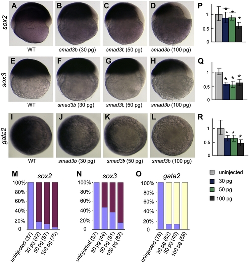
Effects of smad3b overexpression on the expression of early neural and non-neural markers. (A?L) Embryos were injected at the one-cell stage at indicated doses and examined by in situ hybridization for marker gene expression at around 60% epiboly stage. Embryos were positioned laterally with dorsal to the right and animal pole to the top. Note that smad3b overexpression enhanced ventral expression but inhibited dorsal expression of sox2 and sox3. In contrast, smad3b overexpression inhibited expression of gata2. (M?O) Bar graphs showing percentage of embryos with different expression levels of the markers. The number of observed embryos was indicated in the parentheses. For sox2 and sox3, the percentage (red) of embryos with ventral enhancement was calculated. For gata2, the percentage (yellow) of embryos with decreased expression was calculated regardless of decreasing extents. (P?R) Quantitative RT-PCR results of sox2 (P), sox3 (Q) and gata2 (R). See Fig. 1 legend for interpretation. EXPRESSION / LABELING:
|
|
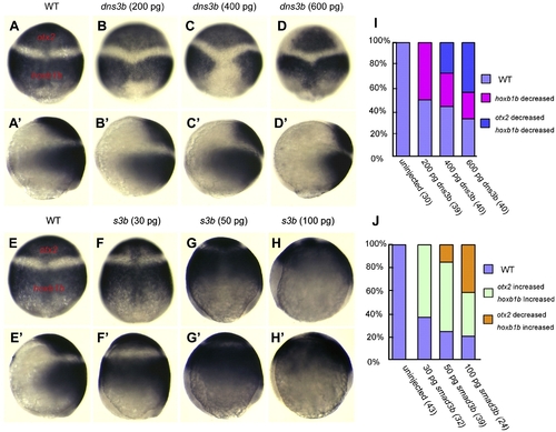
Effects of dnsmad3b or smad3b overexpression on anteroposterior neuroectodermal patterning. Embryos were injected at the one-cell stage with dnsmad3b (dns3b) mRNA (A?D and A′?D′) or smad3b (s3b) mRNA (F?I and F′?I′) at indicated doses, and at around 90% epiboly stage simultaneously examined for the expression of the anterior neural marker otx2 and the posterior neural marker hoxb1b. Embryos were shown in dorsal view (A?H) with animal pole to the top or in lateral view (A′?H′) with animal pole to the top and dorsal to the right. Note that dnsmad3b injection caused both otx2 and hoxb1b domains to shrink from the ventral neuroectoderm but had little effect on the anteroposterior positioning of their boundary. By contrast, smad3b overexpression led to ventral expansion of both otx2 and hoxb1b domains and anterior expansion of the hoxb1b domain. (I, J) Statistical data for dnsmad3b (I) or smad3b (J) injection. The orange color in (J) indicated the proportion of embryos with ventral and anterior expansion of the hoxb1b domain accompanied by anterior reduction of the otx2 domain. The number of observed embryos was indicated in the parentheses. EXPRESSION / LABELING:
|
|
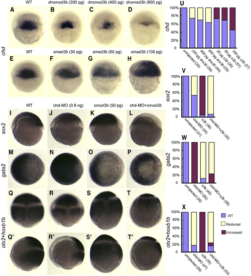
Smad2/3 exert their effects on neural development partially depending on Chd. Embryos were injected at the one-cell stage with an mRNA species or chd-MO as indicated, and examined for the expression of chd (A?H), sox2 (I?L) and gata2 (M?P) at the shield stage as well as for the expression of otx2/hoxb1b at the 90% epiboly stage (Q?T and Q′?T′). Orientations of embryos: dorsal views with animal pole to the top (A?H and Q?T); animal-pole views with dorsal to the right (M?P); and lateral views with dorsal to the right (I?L and Q′?T′). EXPRESSION / LABELING:
|
|
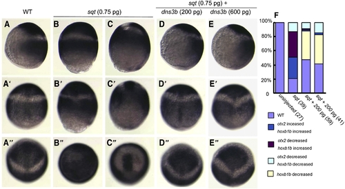
Smad2/3 partially mediate effects of Nodal signal on neural development. Embryos were injected at the one-cell stage with 0.75 pg sqt mRNA alone or together with different doses of dnsmad3b mRNA as indicated. The expression of otx2 and hoxb1bwas simultaneously examined at late gastrulation stages. (A?E) Lateral views with dorsal to the right. (A′?E′) Dorsal views corresponding to those embryos in (A?E). (A″?E″) Anterodorsal views to show the otx2 domain. Overexpression of sqt caused ventral expansion of otx2 and hoxb1b and anterior shift of their boundary at different extents (B, C), which was compromised by co-overexpression of dnsmad3b. EXPRESSION / LABELING:
|
|
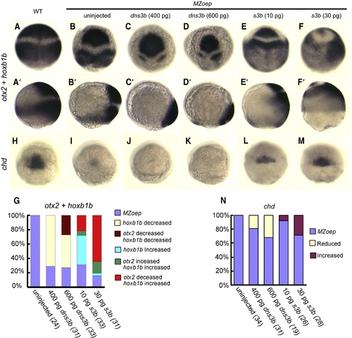
Smad2/3 can exert their effects on neural development in the absence of the organizer. (A, A′ and H) Uninjected wildtype (WT) embryos. (B?F, B′?F′, and I?M) MZoep mutant embryos were injected at the one-cell stage with different doses of dnsmad3b or smad3b mRNA as indicated. Embryos were examined for otx2 and hoxb1b expression at the 90% epiboly stage (A?F and A′?F′) or chd expression at the shield stage (H?M). (A?F and H?M) Dorsal views with animal pole to the top. (A′?F′) Lateral views with dorsal to the right. (G, N) Statistical data for expression of otx2 and hoxb1b (G) or chd (N). EXPRESSION / LABELING:
|
|
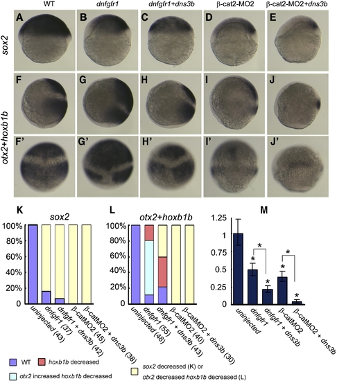
Smad2/3 activities cooperate with Fgf and Wnt signals in neural induction. (A, F and F′) Uninjected wildtype (WT) embryo. (B?E, G?J and G′?J′) Embryos were injected with indicated mRNA species or morpholino at the following doses: 300 pg dnfgfr1 mRNA; 200 pg dnsmad3b mRNA; and 20 ng β-cat2-MO2. (A?E) Lateral views of embryos showed sox2 expression at the shield stage, with dorsal to the right. (F?J and F′?J′) Lateral (F?J) or dorsal (F′?J′) views showed otx2 and hoxb1b expression at late gastrulation stages, with the anterior to the top. (K, L) Statistical data for the percentage of embryos showing altered expression of sox2 (K) and otx2/hoxb1b (L). (M) Quantitative RT-PCR results showing the relative levels of sox2 transcripts at the shield stage after injections with different mRNA species. EXPRESSION / LABELING:
|
|
Effects of Fgf or Wnt signals on neuroectodermal posteriorization can be counteracted by inhibition of Smad2/3 activities. (A, A′, E and E′) Uninjected wildtype (WT) embryos. (B?D, B′?D′, F?H and F′?H′) Embryos were injected with indicated mRNA species at the one-cell stage and examined simultaneously for expression of otx2 and hoxb1b at late gastrulation stages. (A?D and E?H) Lateral views with dorsal to the right. (A′?D′ and E′?H′) Dorsal views with the anterior to the top. (I, J) Statistical data for the percentage of embryos showing altered expression of otx2/hoxb1b. EXPRESSION / LABELING:
|
Reprinted from Developmental Biology, 333(2), Jia, S., Wu, D., Xing, C., and Meng, A., Smad2/3 activities are required for induction and patterning of the neuroectoderm in zebrafish, 273-284, Copyright (2009) with permission from Elsevier. Full text @ Dev. Biol.


