- Title
-
Neuroepithelial cells require fucosylated glycans to guide the migration of vagus motor neuron progenitors in the developing zebrafish hindbrain
- Authors
- Ohata, S., Kinoshita, S., Aoki, R., Tanaka, H., Wada, H., Tsuruoka-Kinoshita, S., Tsuboi, T., Watabe, S., and Okamoto, H.
- Source
- Full text @ Development
|
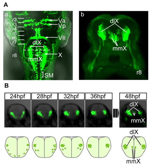
Visualization and live imaging of vagus motor nuclei in isl1:GFP transgenic zebrafish. (A) Fluorescent images of the hindbrain of the isl1:GFP transgenic zebrafish. (a) Dorsal view (rostral towards the top) and (b) cross-section (dorsal towards the top) at the broken line shown in a. (B) Time-lapse imaging of the development of the vagus motor nuclei (upper row), accompanied by the schematic diagrams (lower row; dorsal towards the top). dlX, dorsolateral motor nucleus of the vagus; mmX, medial motor nucleus of the vagus; SM, spinal motor neurons. |
|
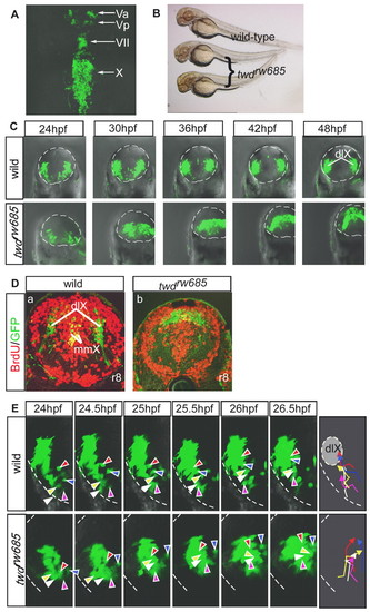
The twdrw685 mutant shows aberrant migration of vagus motor neuron progenitors. (A) Dorsal view of the twdrw685 embryos at 50 hpf (rostral towards the top). (B) Morphology of wild-type and twdrw685 embryos at 2 dpf. (C) Time-lapse imaging of the behavior of the vagus motor neuron progenitors in wild-type (upper row) and twdrw685 (lower row) embryos. The broken line outlines the hindbrain. dlX, dorsolateral motor nucleus of the vagus. (D) Vagus motor neurons and incorporated BrdU at 38 hpf were detected by anti-BrdU (red) and anti-GFP (green) antibodies in wild-type (a) and twdrw685 (b) embryos at 72 hpf (cross-sections; dorsal towards the top). mmX, medial motor nucleus of the vagus. (E) Migratory pathways of five randomly chosen progenitors of the dlX, indicated by different-colored arrowheads. The broken lines show the outline of neural tubes. The lines indicate the trajectories of the individual neurons and correspond to the colors shown in the right-most schematic diagrams. |
|
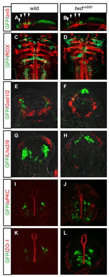
The effect of the twdrw685 mutation is specific to neuronal migration. (A-L) Commissural axons (arrowheads) labeled with zn-5 antibody at 36 hpf (A,B; lateral view, rostral towards the left), reticulospinal neurons retrogradely labeled with rhodamin-dextran (RDX) at 5 dpf (C,D; ventral view, rostral towards the top), Gad1/2-positive neurons at 48 hpf (E,F; cross-sections, dorsal towards the top), Lhx2/9-positive neurons at 48 hpf (G,H; cross-sections, dorsal towards the top), aPKC at 30 hpf (I,J; cross-sections, dorsal towards the top) and ZO-1 at 30 hpf (K,L; cross-sections, dorsal towards the top) are labeled (red) in the wild-type (A,C,E,G,I,K) and twdrw685 mutant (B,D,F,H,J,L) embryos. |
|
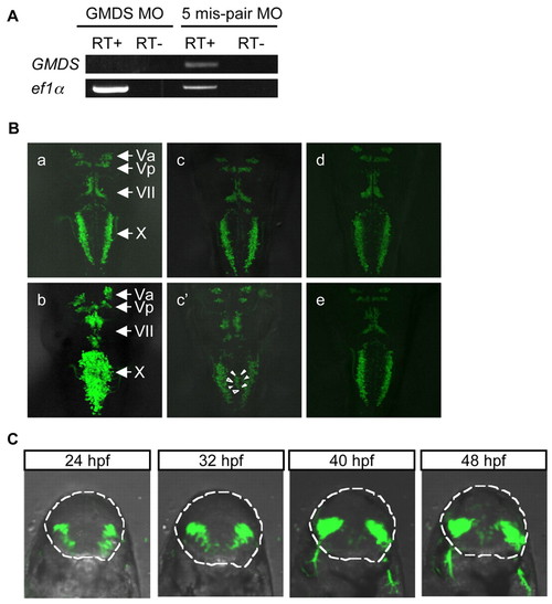
Confirmation of the gmds gene as the twdrw685 locus by knock-down and rescue experiments. (A) The efficacy of the splice-blocking MO against gmds was confirmed by RT-PCR at 32 hpf. Total RNA of gmds morphants and 5-mis-pair control morphants was incubated with (RT+) or without (RT?) reverse transcriptase. Samples were amplified with primers in which the target region of the MO is covered. Therefore, a band is not detected if maturation of gmds mRNA is inhibited by the MO. ef1α; loading control. (B) Dorsal views of the gmds morphant and twdrw685 mutant embryos injected with GMDS mRNAs at 50 hpf (rostral towards the top). Wild-type embryos injected with the 5-mis-pair control MO (a), the gmds MO (b) and S-GMDS mRNA (e). The twdrw685 mutants injected with L-gmds mRNA (c,c′) and S-gmds mRNA (d). Completely rescued (c,d) and partially rescued (c′) mutants. Arrowheads indicate ectopic vagus motor neurons. (C) Time-lapse observation of the migration of vagus motor neuron progenitors in twdrw685 embryos injected with L-gmds mRNA. The broken line shows the outline of the hindbrain. Dorsal is towards the top. |
|
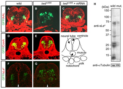
The expression of fucosylated glycans is reduced in twdrw685 embryos. (A-G) Vagus motor neurons and glycans were detected by anti-GFP antibody, biotin-conjugated Aleuria aurantia lectin (AAL; A-C), concanavalin A (ConA; D,E) and anti-sLex (F,G) antibody. Staining of cryosections of wild-type embryos (A,D,F), twdrw685 embryos (B,E,G) and twdrw685 embryos injected with wild-type L-gmds mRNA (C) at 30-34 hpf. Dorsal is towards the top. Arrowheads indicate the ventricular zone (F,G). (H) anti-sLex blot of total lysates from wild-type and twdrw685 mutant embryos. αTubulin was used as a loading control. |
|
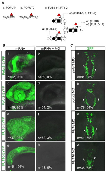
Repression of FUT7-9, FT1, FT2, POFUT1 and POFUT 2 does not phenocopy twdrw685 mutant. (A) Schematic diagram showing the fucosylation sites for POFUT1 (a), POFUT2 (b), FUT4-11 and FT1-2 (c). Red triangle, fucose; diamond, sialic acid; filled circle, galactose; square, GlcNAc; open circle, mannose. (B) (a,c,e,g) Wild-type embryos injected with the mRNAs only; (b,d,f,h) wild-type embryos injected with the mRNAs plus the MOs against the mRNAs. Lateral view at 24 hpf, rostral towards the left. The numbers of embryos examined and the percentage of EYPF-positive embryos are given in the individual pictures. (C) Dorsal view of pofut1 (a), pofut2 (b), FUT8 (c) and FUT10 (d) morphants at 50 hpf (rostral to the top). The numbers of morphants examined and the percentage of morphants with aberrant positioning of the vagus motor neurons lateral to the vagus motor nuclei (a-c, arrowheads) or with fusion of the bilateral vagus motor nuclei (d) are given in the individual pictures. In some FUT10 morphants, aberrant positioning of the vagus motor neurons lateral to the vagus motor nuclei was also observed (d, arrowheads). |
|
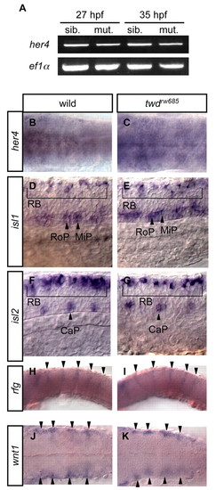
The twdrw685 mutation does not reduce the Notch activity. (A-C) Expression of the Notch target gene her4 was examined by RT-PCR using cDNAs synthesized from the total RNA of the whole body of wild-type and twdrw685 embryos at 27 and 35 hpf (A), and by in situ hybridization of the wild-type (B) and twdrw685 mutant (C) embryos at 24 hpf. Dorsal views, rostral towards the left. (D-G) Rohan-Beard sensory neurons (RB), caudal primary motoneurons (CaP), middle primary motoneurons (MiP) and rostral primary motoneurons (RoP) were visualized by in situ hybridization against isl1 (D,E) or isl2 (F,G) at 25 hpf in wild-type (D,F) and twdrw685 mutant (E,G) embryos. Lateral views, rostral towards the left. (H-K) The rhombomere boundaries were examined by in situ hybridization against radical fringe (rfg; H,I; lateral views, rostral towards the left) or wnt1 (J,K; dorsal views, rostral towards the left) at 24 hpf in wild-type (H,J) and twdrw685 mutant (I,K) embryos. Arrowheads indicate the rhombomere boundaries. EXPRESSION / LABELING:
|
|
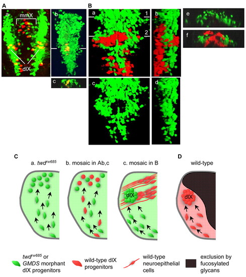
Surrounding neuroepithelial cells regulate the migration of progenitors of the dorsolateral motor nuclei of the vagus (dlX). (A) The wild-type vagus motor neuron progenitors (yellow) were transplanted into wild-type (a) and the gmds morphant (b,c) hindbrain. Dorsal view (a,b; rostral towards the top) and cross-section (c; dorsal towards the top) at the level of the line shown in b. The embryos were observed at 48 hpf. dlX, dorsolateral motor nucleus of the vagus; mmX, medial motor nucleus of the vagus. (B) Wild-type cells (red) were transplanted into the dorsomedial region of the GMDS morphant hindbrain. In this case, the morphant vagus motor neuron progenitors migrated to their correct location (line 2 in a). Dorsal view (a; rostral towards the top) and lateral view (b; rostral towards the top, dorsal towards the right). To observe details of the transplanted embryos, pictures are shown without wild-type cells; c and d correspond to a and b, respectively. Cross-sections of transplanted embryos at the level of lines 1 (e) and 2 (f) in a. The embryos were observed at 48 hpf. (C) Relationship between neural progenitor migration and fucosylation in dlX formation in embryos of the twdrw685 mutant (a), the mosaic in A, parts b,c (b), and the mosaic in B (c). The twdrw685 and GMDS morphant and wild-type dlX progenitors are shown in green and red, respectively. The wild-type neuroepithelial cells are shown in red with the processes. (D) Model to explain how fucosylated glycans regulate the behavior of vagus motor neuron progenitors. There are three steps involved: differentiation near the ventral midline, migration in the dorsolateral direction, and cessation of migration and accumulation of the progenitors at their defined locations. During these developmental steps, dorsomedially distributed fucosylated glycan prevents the dlX progenitors from entering the dorsomedial region of the hindbrain. In the twdrw685 embryos, the progenitors show aberrant migration because this inhibition is lacking. |
|
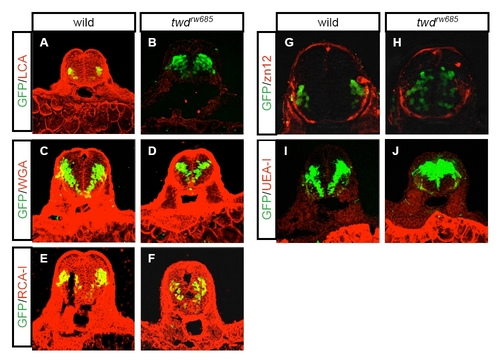
Distribution of various glycans in wild-type and twdrw685 embryos. Vagus motor neurons and glycans are detected by anti-GFP antibody, and the biotin-conjugated lectins or zn12 antibody. Lens culinaris agglutinin (LCA; A,B), wheat-germ agglutinin (WGA; C,D), Ricinus communis agglutinin I (RCA-I; E,F), zn12 antibody (G,H), Ulex europaeus agglutinin I (UEA-I; I,J). Wild-type (A,C,E,G,I) and twdrw685 (B,D,F,H,J) embryos were fixed at 2 dpf and stained. |

