- Title
-
Six3 represses nodal activity to establish early brain asymmetry in zebrafish
- Authors
- Inbal, A., Kim, S.H., Shin, J., and Solnica-Krezel, L.
- Source
- Full text @ Neuron
|
Combined Loss of Six3b/Six7 Function Results in Lack of Eye Tissue and Abnormal Brain Laterality (A) Schematic presentation of normal Six3b and the predicted truncated Six3b protein, encoded by the six3bvu87 allele. (B and C) Live control embryo (B) and a six3b/six7-deficient embryo (B) demonstrating lack of eye tissue. (D and E) Comparable brain patterning in 24 hpf control (D) and six3b/six7-deficient (E) embryos. Forebrain (fb), midbrain-hindbrain boundary (mhb), and hindbrain (hb) are labeled with emx3, pax2a, and egr2b, respectively. The optic stalk (os, arrowheads, pax2a) is reduced in the six3b/six7-deficient embryo. (F and G) Pineal (arrow) and parapineal localization (arrowheads) in control (F) and six3b/six7-deficient (G) embryos. (H and I) Habenular nuclei labeled with lov, in control (H) and six3b/six7-deficient (I) embryos. Control embryos are six3bvu87/+ or six3bvu87/vu87 and present normal phenotypes. R, right; L, left. Anterior is to the left in (B)?(E) and up in (F)?(I). (B)?(E) are lateral views, and (F)?(I) and insets in (B) and (C) are dorsal views. Scale bars, 200 μm. |
|
Brain-Specific Loss of Asymmetry and Excessive Diencephalic Nodal Activation in six3b/six7-Deficient Embryos (A?F) Left-sided expression in control (A, C, and E) and bilateral expression in six3b/six7-deficient embryos (B, D, and F) of lft1 (A and B), pitx2 (C and D), and cyc (E and F). In the LPM, expression remains left-sided (arrows in insets [A?D]). (E?H) The diencephalic domain of Nodal expression and activity is expanded in six3b/six7-deficient embryos (compare [F] to [E] and [H] to [G]). lft1 expression extends outside the flh-positive pineal anlage (H). (I and J) oep expression continues to coincide with the pineal anlage in six3b/six7-deficient embryos (J). Control embryos in all panels are six3bvu87/+ or six3bvu87/vu87 and present normal phenotypes. R, right; L, left. (A?D) Dorsoposterior views; insets show smaller magnification of the same embryo in each panel. (E?J) Dorsal views, anterior is up. All embryos are 21?24s stage. Scale bars, 100 μm. EXPRESSION / LABELING:
|
|
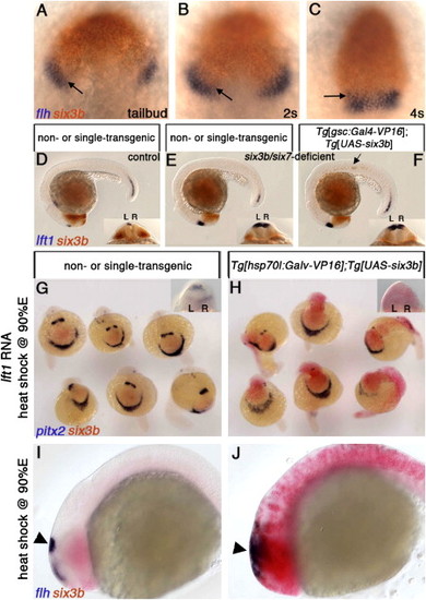
Six3 Function Is Required in the Neuroectoderm to Repress Diencephalic Nodal Activity (A?C) Wild-type embryos at tailbud (10 hpf) (A), 2s (B), and 4s (C) stages, labeled for six3b and flh expression, which marks presumptive epithalamic and dorsal telencephalic tissues. Overlap between six3b and the flh-positive domain is indicated by arrows. (D?F) Embryos from a cross between six3bvu87/+;Tg[gsc:Gal4-VP16]vu160 heterozygous and six3bvu87/+;Tg[UAS-six3b]vu156 heterozygous fish. Control embryo (D) not injected with MOsix7. (E and F) six3b/six7-deficient embryos. Only the embryo in (F) carries both transgenes, as evidenced by ectopic six3b in the notochord (arrow). (G and H) Embryos from a cross between Tg[hsp70l:Gal4-VP16]vu22 and Tg[UAS-six3b]vu156 heterozygous fish that were injected with lft1 RNA and heat-shocked at late gastrulation (H). (I and J) Ubiquitous six3b does not eliminate presumptive pineal tissue (flh-positive domain, arrowheads in [I] and [J]). R, right; L, left. (A?C) Dorsal views, anterior up. (D?F, I, and J) Lateral views, anterior to the left. Insets in (D)-(H) are dorsoposterior views. EXPRESSION / LABELING:
|
|
Six3 Misexpression Can Repress Diencephalic Nodal Activity If Induced by the Beginning of Somitogenesis Tg[hsp70l:Gal4-VP16]vu22;Tg[UAS-six3b]vu156 (B, D, and F) and single or nontransgenic siblings (A, C, and E), heat-shocked at tailbud (10 hpf) (A and B), 4s stage (C and D), or 6s stage (E and F). Arrowheads point at the left-sided dorsal diencephalic domain of Nodal activity represented by pitx2 expression. (G?J) Embryos from a cross between six3bvu87/+;Tg[hsp70l:Gal4-VP16]vu22 heterozygous and six3bvu87/+;Tg[UAS-six3b]vu156 heterozygous fish, which were injected with MOsix7 and heat-shocked at 80% epiboly (80%E) (G and H) or 4s stage (I and J). Insets are dorsoposterior view of the same embryo (A?F) or a representative embryo (G?I). L, left; R, right. Embryos are 22?24s stage. Genotypes in (G), (I), and (J) were inferred from clearly reduced eye size, demonstrating that embryos are six3b/six7 deficient. Because inducing six3b expression by heat shock at 80%E rescues the reduced eye size in six3b/six7-deficient embryos, embryos in (H) were PCR genotyped and the presence of all genotypes at expected ratios was confirmed. EXPRESSION / LABELING:
|
|
Loss of Six3b/Six7 Is Epistatic to Loss of Spaw Function, and a Model for Six3 Function in Regulation of Diencephalic Nodal Activity (A) Control, uninjected embryo. (B) six3bvu87/+ or six3bvu87/vu87 injected with Spaw-MO1 (Long et al., 2003) (MOspaw). lft1 expression is abolished in the diencephalon (arrowhead) and very strongly reduced in the posterior notochord (arrow). (C) six3b/six7-deficient embryo. (D) six3b/six7-deficient embryo injected with Spaw-MO1. Excessive, bilateral diencephalic lft1 expression is not abolished, but lft1 posterior notochord expression is strongly reduced. (E) In clutches comprising approximately 1:1 ratio of six3bvu87/+ and six3bvu87/vu87 embryos, inhibiting Spaw function abolishes lft1 diencephalic expression in 99% of embryos, inhibiting Six7 function causes bilateral lft1 expression in more than 40% of embryos (all six3bvu87/vu87), and inhibiting both Spaw and Six7 results in restoring lft1 expression in 24% of embryos (all six3bvu87/vu87). (F) A model of Six3 function and interaction with Nodal signaling during the establishment of early brain asymmetry. Six3 activity in the anterior neuroectoderm (light blue) and early Nodal signaling, presumably from the axial mesoderm (red), induce by early segmentation repressor/s of Nodal expression (X, dark blue) in the prospective dorsal diencephalon. Later, diencephalic left-sided expression of Nodal is achieved by ipsilateral Spaw, repressing X and activating Nodal. This results in correct epithalamic asymmetries (left-sided parapineal; higher lov levels [gray] in left habenular nucleus). P, pineal; pp, parapineal; lhn, left habenular nucleus; rhn, right habenular nucleus. EXPRESSION / LABELING:
|
|
six3b RNA, but not six3bvu87 RNA, Rescues the Eye Deficiency in six3b/six7-Deficient Embryos Embryos from a cross between six3bvu87/vu87 parents. (A) Uninjected controls. (B) Embryos injected with MOsix7 (six3b/six7-deficient embryos). (C) six3b/six7-deficient embryos injected with 500 pg synthetic six3bvu87 RNA. (D) six3b/six7-deficient embryos injected with 5 pg six3b RNA. This amount of six3b RNA often causes dorsalization. Embryos in top of each panel are representative individuals from the groups below. Scale bar for embryos in top part of each panel is 200 μm. |
|
Prechordal Plate Appears Normal in six3b/six7-Deficient Embryos (A, C, E, G) Control, uninjected embryos (six3bvu87/+ or six3bvu87/vu87), stained with prechordal plate markers at the end of gastrulation (9.5-10 hpf). (B, D, F, H) Representative sibling embryos injected with MOsix7. EXPRESSION / LABELING:
|


