- Title
-
Expression pattern of zebrafish tcf7 suggests unexplored domains of Wnt/beta-catenin activity
- Authors
- Veien, E.S., Grierson, M.J., Saund, R.S., and Dorsky, R.I.
- Source
- Full text @ Dev. Dyn.
|
Clustal alignment of predicted amino acid sequences of Tcf7 in zebrafish (Danio rerio, D.r.), Xenopus laevis (X.l.), mouse (Mus musculus, M.m.), and human (Homo sapiens, H.s.). The amino acid sequences are 53%, 49%, and 46% identical to zebrafish Tcf7 B isoform, respectively. A: Amino acid alignment up to the point of alternative C-termini (amino acid #400). Highlighted are the amino terminal β-catenin binding domain, the putative Groucho binding domain, and the C-terminal HMG box. The mouse form shown does not include the Groucho binding domain; however, other splice forms do. B: Amino acid alignments of C-terminal Tcf7 splice forms corresponding to the human B, C, and D isoforms share significant amino acid identity. |
|
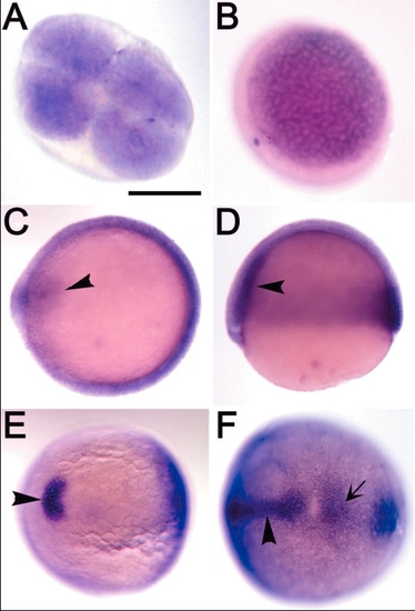
In situ hybridization analysis of tcf7 expression from four-cell to bud stage. A: Animal pole view. Ubiquitous maternal expression at four-cell stage. B: Animal pole view. Ubiquitous expression at dome stage. C: At 60% epiboly. Specific expression in shield and prechordal mesoderm (arrowhead) as well as ubiquitous expression in germ ring. Dorsal to left. D: Lateral view at 60% epiboly shows higher level of expression in the hypoblast (arrowhead). Dorsal to left. E,F: Bud stage. E: Expression in the prechordal plate (arrowhead). F: The arrow indicates expression in the tail bud, and the arrowhead indicates expression in notochord. Posterior view, dorsal to left. Scale bar = 300 μm in A (applies to A-F). |
|
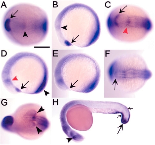
Expression of zebrafish tcf7 during somitogenesis. A,B: The five-somite stage. Arrows indicate expression in prechordal plate, and arrowheads indicate expression in diencephalon/mesencephalon boundary. A: Dorsal view, anterior to left. B: Lateral view, anterior to left. C,D: The eight-somite stage. Specific expression seen in diencephalon/mesencephalon boundary (red arrowheads), polster (arrows), and tail bud (black arrowhead). C: Dorsal view, anterior to left. D: Lateral view, anterior to left. E,F: The 12-somite stage. Expression of tcf7 is absent in polster (arrows). E: Lateral view, anterior to left. F: Dorsal view, anterior to left. G: The 15-somite stage showing expression in dorsal retina (arrowheads). Frontal view. H: Embryo at 24 hr postfertilization (hpf) showing expression in dorsal retina (arrowhead), tail bud (large arrow), and median fin fold (small arrows). Lateral view, anterior to left. Scale bar in A = 300 μm in A-G, 500 μm in H. EXPRESSION / LABELING:
|
|
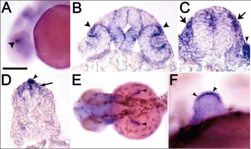
Expression of tcf7 in specific regions at 24-36 hours postfertilization (hpf). A: Expression in dorsal retina (arrowhead) at 24 hpf. Lateral view, anterior to left. B: Coronal section through the diencephalon at 24 hpf. Higher level of expression in margin of the dorsal retina (arrowheads) and ventral diencephalon. C: Coronal section through the anterior hindbrain at 24 hpf. Expression in dorsal hindbrain, neural crest (arrows), and otic vesicle (arrowhead). D: Coronal section through spinal cord at 24 hpf. tcf7 expression is seen in dorsal spinal cord (arrow) and median fin fold (arrowhead). E,F: Expression in pectoral fin apical fold ectoderm at 36 hpf. E: Dorsal view of fin buds, indicated by arrowheads. F: Lateral view of pectoral fin bud showing high level of expression in basal layer ectodermal cells (arrowheads). Apical cells are unstained. Scale bar in A = 300 μm in A,E, 175 μm in B-D, 100 μm in F. |

Unillustrated author statements |


