- Title
-
Zebrafish smoothened functions in ventral neural tube specification and axon tract formation
- Authors
- Varga, Z.M., Amores, A., Lewis, K.E., Yan, Y.-L., Postlethwait, J.H., Eisen, J.S., and Westerfield, M.
- Source
- Full text @ Development
|
The smu mutant phenotype is due to mutations in smoothened. (A) 24 h wild type (top); 24 h smub641 mutant (bottom). (B) In smub641 mutants (bottom), floor plate was poorly differentiated, somites lacked a horizontal myoseptum and were U-shaped compared with wild type (top). (A,B) Side views, dorsal is towards top, anterior is towards left. Scale bar: 300 μm in A; 50 μm in B. (C) Phylogenetic analysis. Statistical robustness was estimated by bootstrap, numbers indicate the percent of times each node was obtained. Danio rerio (Dre), Drosophila melanogaster (Dme, P91682), Homo sapiens (Hsa, NP_005622), Gallus gallus (Gga, O42224), Mus musculus (Mmu, P56726) and Rattus norvegicus (Rno, NP_036939). (D) Syntenic relationships. Conserved syntenies to Hsa 7q32.3 indicate that smoothened is a zebrafish ortholog of mammalian smoothened. (E) Mapping. Diagram of LG4 showing positions of smoothened (smoh) gene and smu mutations. (F) Model of protein encoded by smoothened. The red (smub641) and green (smub577) dots indicate amino acids altered in the two alleles. A seven-pass transmembrane protein is the predicted structure of zebrafish Smoothened (TMHMM1.0 software, Center Biol. Sequence Analysis, Department Biotech., Tech. University of Denmark). (G) Mutations. Point mutations in smub641 (red) and smub577 (green). PHENOTYPE:
|
|
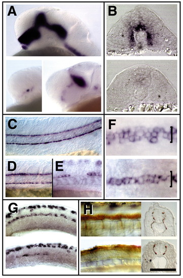
smu mutants have defective Hh signaling. (A) In 24 h smub641 mutants (bottom left), nkx2.2 gene expression was reduced compared with wild type (top); nkx2.2 gene expression was less severely affected in smub577 mutants (bottom right). (B) At the 16-somite stage, patched1 expression was essentially absent from smub641 mutants (bottom) compared with wild type (top). (C-E) At 24 h, floor plate and hypochord were present in both mutant (D) and wild-type (C) embryos labeled for col2a1 (Yan et al., 1995). By 27 h (E) gaps appeared in smu mutant floor plate indicated by shh expression. (F) At 20-somite stage, mutants (bottom) lacked lateral floor plate compared with wild type (top), indicated by foxa2. (G) At the 18-somite stage, islet1-expressing primary motoneurons were absent from tail and posterior trunk, and were reduced in anterior spinal cord in smub641 mutants (bottom) compared with wild type (top). (H) In smub641 mutants (bottom), secondary motoneurons were absent at 24 h, indicated by DM-GRASP labeling. (A,C-E,G,H) Side views, dorsal is towards the top, anterior is towards the left. (B,H, insets) Transverse sections of anterior trunk, dorsal is towards the top. (F) Dorsal views, anterior is towards the left. Scale bar:120 Ám (A, G); 110 Ám (B,H); 100 Ám (C-E); 50 Ám (F); 82 Ám (H, insets). EXPRESSION / LABELING:
PHENOTYPE:
|
|
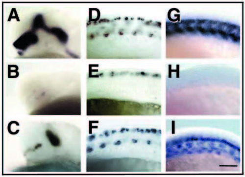
The smu mutant phenotype is rescued by injection of smoothened RNA. (A,B) At 24 h, smub641 embryos (B) had almost no nkx2.2 expression compared with wild type (A). (C) Injection of 50 pg smoothened RNA at the two-cell stage partially rescued nkx2.2 expression. (D,E) At 18 h, smub641 embryos (E) lacked islet1-expressing primary motoneurons in posterior trunk and tail compared with wild type (D). (F) Injection of 100 pg smoothened RNA at the one-cell stage completely rescued primary motoneuron phenotype in smub641 mutants. (G,H) At 24 h, smub641 embryos (H) had almost no patched1 expression compared with wild type (G). (I) Injection of 50 pg smoothened RNA at the two-cell stage almost completely rescued patched1 expression. Side views, dorsal towards the top, anterior towards the left. Scale bar: 75 μm in A-C; 50 μm in D-F; 62 μm in G-I. EXPRESSION / LABELING:
|
|
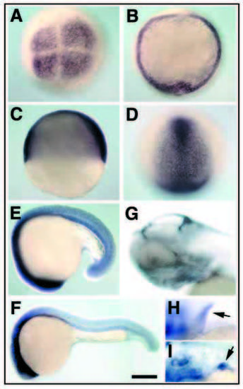
smoothened is expressed both maternally and zygotically. (A) Maternal smoothened mRNA present at four-cell stage. (B,C) smoothened mRNA present throughout the embryo at shield (B) and 60% epiboly (C). (D) smoothened expression was downregulated in the non-neural ectoderm at tailbud stage. (E,F) smoothened was expressed at higher levels in the head than the rest of the body at 18- somites (E) and 24 h (F). (G) By 2 d, smoothened expression in the head was confined to dorsal brain nuclei and jaw cartilages. (H,I) At 2 d, smoothened was also expressed in the pectoral fin of wild-type (H) and smub641 mutant (I) embryos. (A,B,D) Animal pole views. (B,D) Anterior is towards the top. (C,E-I) Side views, dorsal towards the top, anterior towards the left. Scale bar: 200 μm in A-E; 250 μm in F; 115 μm in G; 90 μm in H,I. |
|
smu mutant embryos have residual Hedgehog function provided by maternal Smoothened. shh mRNA increases expression of nkx2.2 after injection into wild-type (B) and smu mutant (D) embryos compared with uninjected wild-type (A) or smu mutants (C). Injection of shh and twhh morpholinos into smu mutants decreases residual nkx2.2 expression (F) compared with uninjected mutants (E). Side views, anterior towards the left; (A-D) two-somite stage; (E, F) Prim-6 stage. Scale bar: 100 μm. |
|
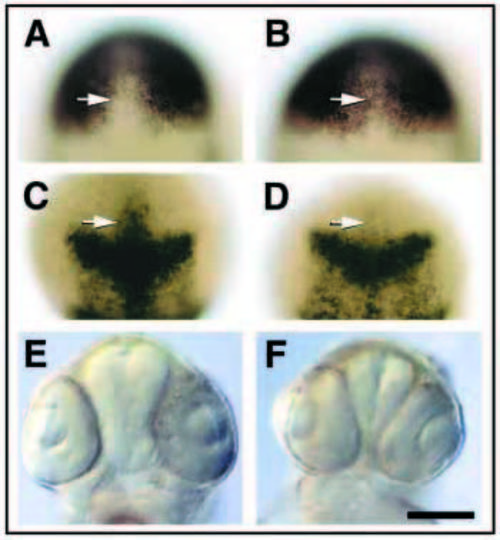
Hh signaling patterns the midline of the anterior neural plate. (A,B) expression of zic1 at tailbud stage in wild-type (A) and smub641 mutant (B) embryos. (C,D) expression of foxb1.2 at tailbud stage in wild-type (C) and smub641 mutant (D) embryos. Arrows indicate location of hypothalamic precursors demarcated by foxb1.2. (E, F) Head morphology of wild-type (E) and smub641 mutant (F) embryos. In 48 h smu mutants, the hypothalamus was reduced in size, consistent with reduced foxb1.2 expression in neural plate precursors and subsequent progressive loss of hypothalamic tissue. (A-D) Dorsal views of prospective head region, anterior towards top. (E,F) Ventral views, anterior towards top. Scale bar: 200 μm in A-D; 135 μm in E,F. |
|
smu mutations disrupt dorsoventral forebrain and retinal patterning, optic stalk formation and pituitary specification. (A) Expression of emx1 in wild-type (top) and smub641 mutant (bottom) embryos. emx1 expression expanded into ventral regions. (B) Expression of dlx2 in wild-type (top) and smub641 mutant (bottom) embryos. Ventral dlx2 expression was reduced in forebrain at the level of anterior and post-optic commissures; dorsal telencephalic expression was expanded. (C) Expression of pax2a in wild-type (top) and smub641 mutant (bottom) embryos. In smu mutants, pax2a expression was lost in optic stalk and ectopic (arrow) in hypothalamus. (D) Expression of pax6a in wild-type (top) and smub641 mutant (bottom) embryos. In smu mutants, pax6a expression was strongly reduced in thalamus. (E) Expression of pax6a expanded in ventral retina of smu mutants (bottom) compared with wild type (top). (F) Expression of pax2a in optic stalk (top, wild type) was lost in smu mutants (middle) and diencephalic cells expressed pax2a ectopically (arrow). An ectopic lens (arrow) developed in smu mutants, as shown in more ventral focal plane (bottom). (G) smu mutations affect specification of the pituitary. The anterior pituitary expressed lim3 in wild-type embryos (top). In place of the pituitary, an ectopic lens (arrow) formed in smu mutants (bottom). (H) Lens fiber cells differentiated in wild type (top), smu mutant ectopic (middle) and retinal lenses (middle, bottom) indicated by zl-1 (red). (A-D) Side views, dorsal towards the top, anterior towards left. (E,F) Dorsal view, anterior towards top. (G,H) Anterior view, dorsal towards top. 24 h. Scale bar in D: 100 μm in A-D; 40 μm in E; 50 μm in F (top); 25 μm in F (bottom); 60 μm in G; in H, 50 μm. |
|
smu mutations disrupt formation of forebrain axon tracts and commissures. (A,B) An optic chiasm (arrow) was formed in wildtype (A) but not smu mutant (B) embryos. In smu mutants (D), anterior commissure axons (arrows) projected into hypothalamus (h), optic chiasm was absent and supra-optic tract axons (S) extended toward the ventral midline (compare with wild type in C). (E-H) Expression of netrin1 is altered in smu mutant embryos. In smu mutant embryos (F) netrin1 expression is upregulated in the midline of the neural tube at the level of the post optic commissure (E,F; arrows) and near the presumptive zona limitans intrathalamica (E,F; arrowheads). The netrin1 expression domain close to the presumptive exit point of ganglion cell axons from the retina (G, arrow) is absent in smu mutants (H, arrow). Ac, anterior commissure; h, hypothalamus; Oc, optic chiasm; P, post-optic commissure; S, supra-optic tract; T, telencephalon; Tp, tract of the post-optic commissure. (A,B) Ventral views, dorsal towards the top, DMGRASP label, 36 h. (C-F) Side views, dorsal towards the top, anterior towards the left, 48 h; (C,D) acetylated tubulin label. (G,H) Dorsal views, anterior towards left. Scale bar: 100 μm in A,B; 22.5 μm in C,D; 85 μm in E-H. |


