- Title
-
Functional analysis of zebrafish GDNF
- Authors
- Shepherd, I.T., Beattie, C.E., and Raible, D.W.
- Source
- Full text @ Dev. Biol.
|
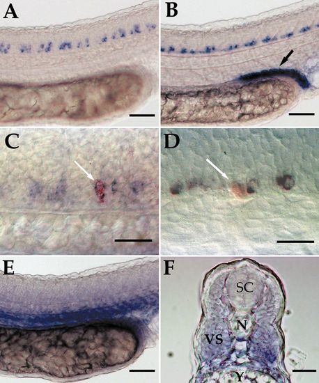
Expression of zebrafish GDNF, GFRα1b, and RET genes in the trunk of 24 hpf embryos (A and B). Low power lateral views of the trunks of 24 hpf embryos hybridized with riboprobes for GFRα1b (A) and RET (B). (C, D) High power lateral views of whole-mount 24 hpf embryos hybridized with GFRα1b riboprobe (blue reaction product) that had been injected with lysinated fluorescence dextran (red reaction product) to identify CaP (C) and MiP (D) PMN prior to in situ hybridization. Low power lateral view of the trunk of 24 hpf embryos hybridized with riboprobes for GDNF (E). Transverse section taken through a 24 hpf embryo hybridized with probe for GDNF at the level of somite 14. SC, spinal cord; N, notochord; VS, ventral somite; Y, yolk. Arrow in (B) indicates pronephric duct. Arrows in (C) and (D) indicate the identified PMN. In (A?E) rostral is to the left. Scale bars: (A), (B), (E), and (F), 50 μm; (C) and (F), 25 μm. EXPRESSION / LABELING:
|
|
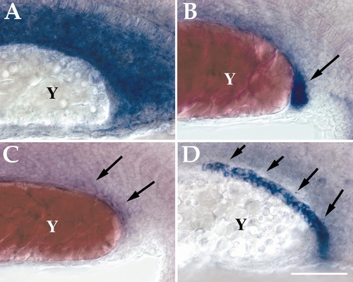
Pronephric duct and mesenchyme mRNA expression of zebrafish GDNF, GFRα1, and RET at 20 hpf. (A?D) Lateral view of the ventral half of the trunk of 18 hpf embryos from somite 10?18 that have been hybridized with riboprobes for zebrafish GDNF (A), GFRα1a (B), GFRα1b (C), and RET (D) messages. Y, yolk sac extension. Arrow in (B) indicates the posterior part of the pronephric duct. Arrows in (C) indicate weak GFRα1b expression in the pronephric duct. Arrows in (D) indicate pronephric duct. Rostral is to the left. Scale bar: 50 μm. EXPRESSION / LABELING:
|
|
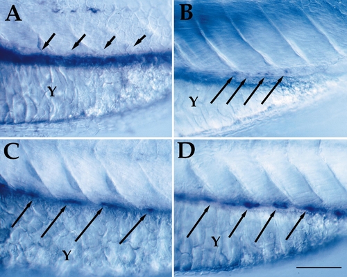
Enteric nervous system and gut endoderm mRNA expression of zebrafish GDNF, GFRα1, and RET at 72 hpf. (A?D) Lateral view of the trunk of 72 hpf embryos from somites 9?13 that have been hybridized with GDNF (A), GFRα1a (B), GFRα1b (C), and RET (D) riboprobes. Y, yolk sac extension. Arrows in (A) indicate gut endoderm. Arrows in (B?D) indicate enteric neuron processors in the gut endoderm. Rostral is to the left. Scale bar: 50 μm. EXPRESSION / LABELING:
|
|
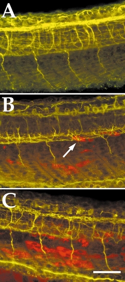
Focal overexpression of GDNF in zebrafish somitic muscle cells causes PMN axon pathfinding errors. (A?C) Lateral view of the trunks of 24 hpf antiacetlyated tubulin antibody stained control (A), focal GDNF overexpressing embryos (B), and uniform GDNF overexpressing embryos (C). Red cells in (B) and (C) express the muscle actin promoter: GDNF DNA construct as revealed by whole-mount in situ hybridization with a riboprobe against a 3′ UTR tag contained in the injected DNA construct. Arrow in (B) indicates a stalled PMN axon projection associated with a GDNF overexpressing cell. Scale bar: 50 μm. |
|
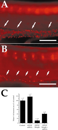
GDNF antisense morpholino injections cause a loss of enteric neurons in zebrafish embryos. Lateral view of the trunks of 96 hpf embryos whole-mount stained with anti-Hu antibody. (A) Control embryo. (B) GDNF morpholino-injected embryo. (C) A bar graph showing the number of enteric neurons in control; GDNF RNA injected; GDNF morpholino injected; and GDNF morpholino plus GDNF RNA injected embryos as a percentage of control. The error bars in (C) indicate 95% confidence interval. Arrows in (A) and (B) indicate gut endoderm. Rostral is to the left in (A) and (B). Scale bars: 100 μm. PHENOTYPE:
|

Unillustrated author statements EXPRESSION / LABELING:
|
Reprinted from Developmental Biology, 231(2), Shepherd, I.T., Beattie, C.E., and Raible, D.W., Functional analysis of zebrafish GDNF, 420-435, Copyright (2001) with permission from Elsevier. Full text @ Dev. Biol.

