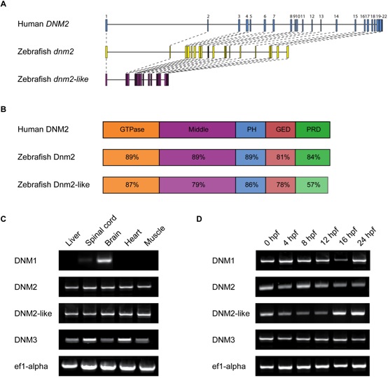
Fig. 2 Structure and expression of dnm2 and dnm2-like.(A) Molecular intron-exon organization of human DNM2, zebrafish dnm2 and zebrafish dnm2-like. (B) Protein structure of zebrafish Dnm2 and Dnm2-like compared to human DNM2. Percent identity between zebrafish and human protein domains was calculated using BLASTP. PH, pleckstrin homology domain; GED, GTPase effector domain; PRD, proline-rich domain. (C) RT-PCR was used to assay spatial expression levels of dnm2 and dnm2-like in tissues isolated from adult zebrafish. Primers for ef1α were used as an internal control. (D) RT-PCR was used to assay temporal expression levels of dnm2 and dnm2-like between 0 hpf and 24 hpf. All classical dynamins appear to be deposited as maternal mRNAs and expressed throughout early development.
Image
Figure Caption
Figure Data
Acknowledgments
This image is the copyrighted work of the attributed author or publisher, and
ZFIN has permission only to display this image to its users.
Additional permissions should be obtained from the applicable author or publisher of the image.
Full text @ PLoS One

