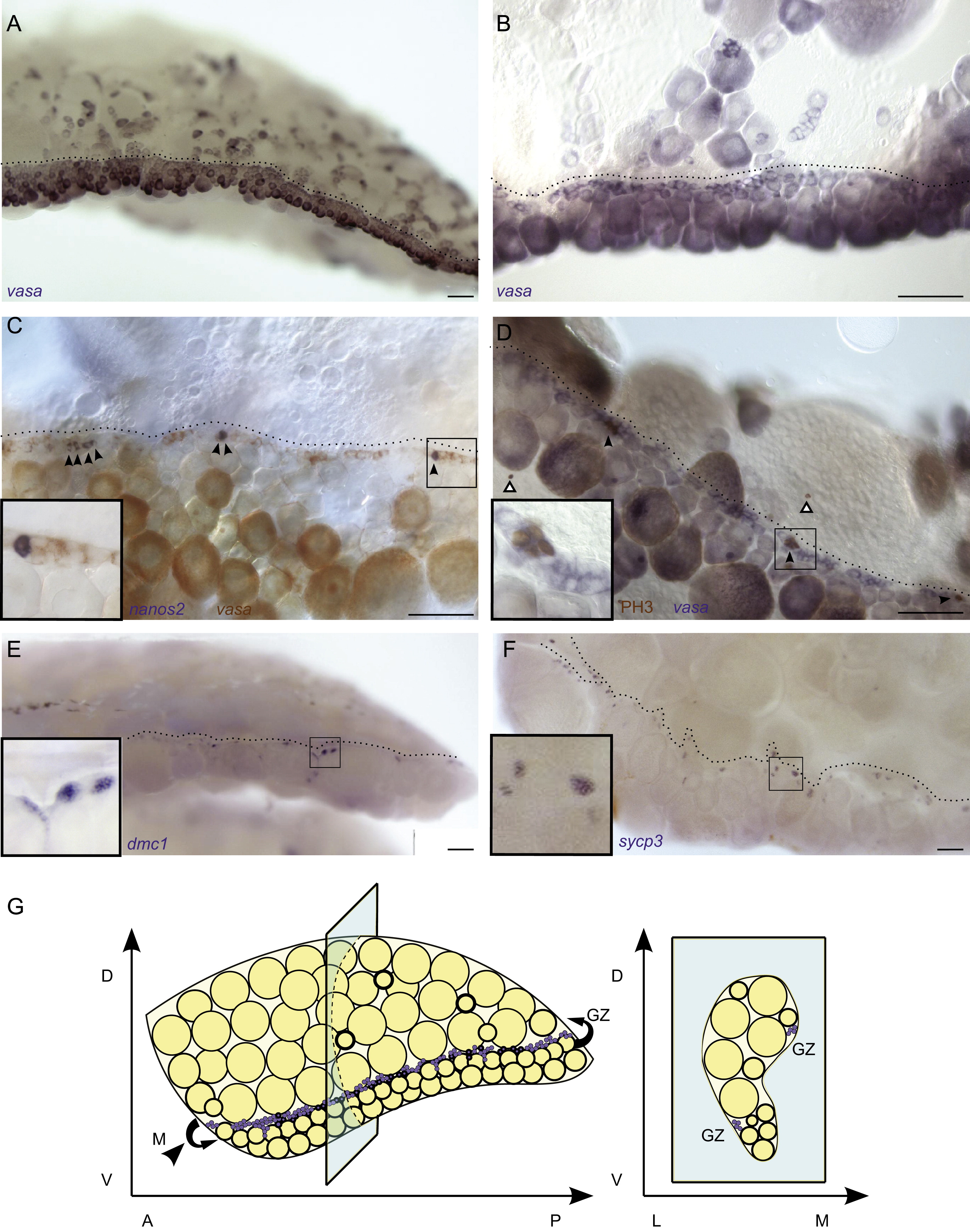
Fig. 4 The germinal zone is the (A and B) vasa RNA in situ hybridization demonstrates that the <20 μm germ cell population in 3 mpf adult ovaries localizes to the germinal zone (dotted line) which is readily identifiable at low magnification (A), and higher magnification (B). (C) Two color RNA in situ hybridization shows that nanos2-expressing cells (blue, indicated by arrowheads) co-localize with the vasa-expressing <20 μm cells (red) in the germinal zone. (D) RNA in situ and immunohistochemistry co-staining reveal that all mitotic germ cells (brown PH3+ and blue vasa+, indicated by black arrowhead) are located in the germinal zone. White arrowheads indicate PH3+vasa- somatic cells. (E and F) dmc1+ cells (blue in E), and sycp3+ cells (blue in F) localize to the germinal zone. Insets in C?F show higher magnification of boxed region. All images are from whole mount preparations. (G) Three-dimensional illustration of an adult ovary. <20 μm cells are indicated in purple. A-anterior, P-posterior, D-dorsal, V-ventral, L-lateral, M-medial and GZ-germinal zone. Scale bars: 100 μm (A, E and F), 50 μm (B?D).
Reprinted from Developmental Biology, 374(2), Beer, R.L., and Draper, B.W., nanos3 maintains germline stem cells and expression of the conserved germline stem cell gene nanos2 in the zebrafish ovary, 308-318, Copyright (2013) with permission from Elsevier. Full text @ Dev. Biol.

