
Fig. 8
- ID
- ZDB-FIG-241202-28
- Publication
- Wang et al., 2024 - Planar cell polarity zebrafish models of congenital scoliosis reveal novel underlying defects in notochord morphogenesis
- Other Figures
- All Figure Page
- Back to All Figure Page
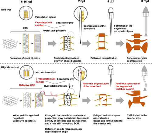
Proposed model for the pathogenic mechanism(s) underlying CVM in MZptk7a mutants. In wild-type zebrafish embryos, starting from the shield stage (?6 hpf), mesodermal cells undergo C&E to form a typical stack of coin-like notochord in the dorsal midline just before the beginning of the vacuolation process (?16 hpf). As notochord cells vacuolate, they are resisted by the notochord sheath, thereby increasing the notochord internal pressure and stiffness, and enabling the notochord to elongate and straighten. At later stages, the notochord provides a scaffold for the patterned spine segmentation. In MZptk7a mutants, a defective C&E leads to a wider and disorganized notochord that would lead to excessive apoptosis around the initiation time of the vacuolation process and a decrease in the number of vacuolated cells. This would cause a decrease in the notochord pressure and a change in the mechanical properties of the notochord, as manifested by a wavy notochord, a decrease in the density of caveolae and desmosomes, and a less stiff notochord ECM. These cellular and mechanical changes would, in turn, lead to notochord kinks, spine mis-segmentation and CVMs limited to the anterior axis. Notochord defects in MZptk7a mutants were accompanied by a wider chevron angle and defects in somite development. C&E, convergence and extension; mpf, months postfertilization |