
Fig. 3
- ID
- ZDB-FIG-241202-23
- Publication
- Wang et al., 2024 - Planar cell polarity zebrafish models of congenital scoliosis reveal novel underlying defects in notochord morphogenesis
- Other Figures
- All Figure Page
- Back to All Figure Page
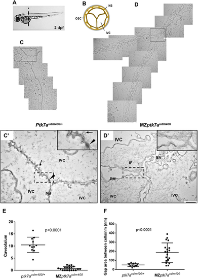
Transmission electron microscopy reveals ultrastructural abnormalities of the notochord vacuolated region in MZptk7audm400 mutants. (A) The level of the transverse cross-sections of 2 dpf embryos for TEM studies is shown. (B) A representative diagram of a transverse section of the notochord. (C-D?) Overlapping ultrastructure images of the junction between two neighboring inner vacuolated cells from MZptk7audm400 embryos (D) and their ptk7audm400/+ siblings (C). C? and D? show higher magnification views of the boxed regions in C and D, respectively. Arrow and arrowhead indicate caveolae and desmosome, respectively. MZptk7audm400 mutants show severely loosened inter-vacuolated cell junctions with an abnormal accumulation of IF and of extracellular vesicles as well a severe reduction in the number of caveolae with an altered morphology in the plasma membrane of the vacuolated cells. Insets show higher magnifications of the boxed areas. Scale bars: 800 nm (C,D); 400 nm (C?,D?). (E) Quantification of the ?normal? caveolae in MZptk7audm400 (3 embryos, 11 images) and their ptk7audm400/+ siblings (3 embryos, 19 images). (F) Quantification of the gap between adjacent vacuolated cells after membrane length standardization. P-values were calculated using two-tailed Mann?Whitney U-test. EV, extracellular vesicles; IF, intermediate filament; IVC, inner vacuolated cell; NS, notochord sheath; OSC, outer sheath cell; PM, plasma membrane. |