
Fig. 3
- ID
- ZDB-FIG-240910-55
- Publication
- Burkhalter et al., 2024 - Cilia defects upon loss of WDR4 are linked to proteasomal hyperactivity and ubiquitin shortage
- Other Figures
- All Figure Page
- Back to All Figure Page
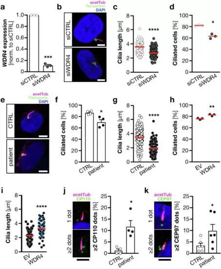
Cilia defects in the absence of WDR4 in human fibroblasts.a siRNA transfection of human fibroblasts results in reduced WDR4 expression. qPCR normalized to housekeeping gene (SDHA) and to siCTRL transfected cells. n = 3. ***p = 0.0003, paired t-test. b Immunofluorescence of cilia in fibroblasts transfected with control or WDR4 siRNA. Magenta: acetylated tubulin (cilia); green: pericentrin (PCNT, centrioles). Scale bar: 5 �m. c Cilia are shorter upon WDR4 KD. n = 3 experiments with 95?99 cilia in total. ****p = 0.0001. Welch?s t test. d WDR4 KD cells have fewer cilia. n = 3 experiments. *p = 0.0108. Unpaired, Welch?s t test. e Immunofluorescence of control and patient-derived primary fibroblasts. Staining as in (a). Scale bar: 5 �m. f Wdr4 patient fibroblasts have fewer cilia. n = 4 experiments. *p = 0.0255. Welch?s t test. g WDR4 patient fibroblasts have shorter cilia. n = 4 experiments with 108?174 cilia in total. ****p < 0.0001. Mann?Whitney test. h Nucleofection of WDR4 patient fibroblasts with a plasmid encoding human WDR4 rescues the percentage of ciliated cells. n = 3 experiments. **p = 0.01. Welch?s t test. i Reconstitution with WDR4 restores cilium length in patient fibroblasts. n = 3 experiments with 99 and 100 cilia. ****p < 0.0001. Welch?s t test. j Wdr4 patient cells show increased frequency of aberrant CP110 staining (green). n = 4 experiments with 407 and 411 cells in total. *p = 0.0455. Paired t-test. Mean + SEM. Scale bar: 3 �m. k Wdr4 patient cells show increased frequency of aberrant CEP97 staining (green). n = 6 experiments with 625 and 617 cells in total. *p = 0.0342. Paired t-test. Mean + SEM. Scale bar: 3 �m. Red line indicates median. |