|
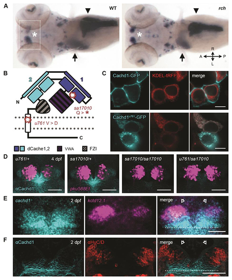
cachd1 mutants show bilaterally symmetrical, double-left habenulae. (A) Dorsal views of whole-mount 5 dpf wild-type sibling and rorschach (rch/u761) mutant larvae showing expression of an asymmetric habenular marker (kctd12.1, indicated with asterisk; box indicates approximate epithalamic region) and markers for liver (selenop2, indicated with arrow), pancreas (prss1, indicated with arrowhead), and ventral retina (aldh1a3). (B) Schematic of Cachd1 protein: two dCache domains (cyan and dark blue), a VWA domain (purple stripes), a FZD interaction domain (FZI; gray stripes), a transmembrane domain (white), and an unstructured cytoplasmic tail. Residues affected in sa17010 and u761 alleles are marked in red at approximate positions in primary sequence. (C) Fluorescence images of transfected HEK293T cells expressing constructs encoding EGFP-tagged wild-type (top; cyan) or rch/u761 mutant Cachd1 (bottom; cyan) and KDEL-tRFP (red) to mark the endoplasmic reticulum. Scale bar, 10 μm. (D) Dorsal views of brains of dissected 4 dpf transgenic siblings from a complementation cross of sa17010 and u761 alleles, stained with antibody to Cachd1 (cyan). The Et(gata2a:EGFP)pku588 (pku588Et) transgene is expressed in dHbL neurons (magenta). (E) Dorsal view of 2 dpf habenulae after double fluorescent in situ hybridization with cachd1 antisense riboprobe (cyan) and the dHbL marker kctd12.1 (magenta). (F) Dorsal views of 2 dpf habenulae after immunohistochemistry with antibody to Cachd1 (cyan) co-stained with antibody to HuC/D to mark differentiating neurons (red). The dotted lines in (E) and (F) indicate the approximate position of the posterior commissure; open arrowheads indicate the dorsal habenulae. Shown are maximum projections of [(D) and (E)] confocal z-stacks or (F) single confocal slice. Scale bars, 50 μm.
|

