
Fig. 1
- ID
- ZDB-FIG-231215-9
- Publication
- Zhang et al., 2022 - Blf and drl cluster synergistically regulate cell fate commitment during zebrafish primitive hematopoiesis
- Other Figures
- All Figure Page
- Back to All Figure Page
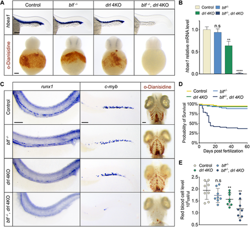
Simultaneous depletion of blf and the drl cluster leads to severe erythrocyte aplasia. (A) Top: RNA in situ hybridization of hbae1 expression in primitive erythrocytes of 23 hpf embryos. Bottom: Whole-mount o-dianisidine staining of 36 hpf embryos. (B) Relative expression levels of hbae1 in 23 hpf embryos. Data are mean�s.e.m. (C) Left: RNA in situ hybridization of runx1 expression in the dorsal aorta of 36 hpf embryos. Middle: RNA in situ hybridization of c-myb expression in the caudal hematopoietic tissue of 3 dpf embryos. Right: Whole-mount o-dianisidine staining of 7 dpf embryos; ventral views. (D) Representative Kaplan?Meier plot for wild-type, blf?/?, drl 4KO and blf?/?; drl 4KO fish from one of three independent experiments. Eighty animals of each genotype were followed. (E) Red blood cell levels in peripheral blood from adult fish; data are mean�s.e.m. Circles represent individual samples. In B and E, n.s, not significant, **P<0.01, ****P<0.0001. Scale bars: 100 ?m. |