
Fig. 3
- ID
- ZDB-FIG-231215-11
- Publication
- Zhang et al., 2022 - Blf and drl cluster synergistically regulate cell fate commitment during zebrafish primitive hematopoiesis
- Other Figures
- All Figure Page
- Back to All Figure Page
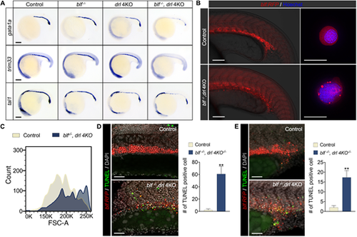
blf and drl cluster genes are essential for establishing the erythroid cell differentiation program. (A) RNA in situ hybridization of gata1a, trim33 and tal1 expression in 18-somite embryos. Lateral view with head to left. For gata1a WISH, n=37, 35, 33 or 36. For trim33 WISH, n=40, 38, 37 or 32. For tal1 WISH, n=31, 25, 40 or 37. Listed sample sizes relate to each group shown from top to bottom. Scale bars: 100 ?m. (B) Fluorescence micrographs of control (blf+/?) and blf?/?; drl 4KO embryos and sorted RFP+ cells at 22 hpf. Scale bars: 200 ?m (left); 10 ?m (right). (C) Histograms of forward scatter showing approximate cell size distributions of the sorted RFP+ cells. (D,E) Left: Cell death in the intermediate cell mass (D) and in EMPs (E) assessed with TUNEL staining at 22 hpf. Scale bars: 100 ?m. Right: Quantification of TUNEL-positive cells counted on a 100 ?m�250 ?m field for inner cell mass and 125 ?m�125 ?m field for EMPs. Data are mean�s.e.m. **P<0.01. Data were collected for 8-12 samples per experiment group. |