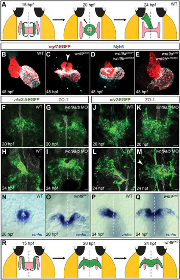
The wnt9a/b paralogous genes are essential for myocardial morphogenesis. (A) Model depicting the zebrafish embryonic wild-type heart during stages of cardiac cone formation and leftward jogging. At 15 hpf, bilateral populations of cardiomyocyte progenitor cells start converging towards the embryonic midline (black arrows). After 20 hpf, endocardial and myocardial progenitor cells initiate unilateral migrations towards the left, which causes a tilting of the heart cone and an elongation of the heart tube. (B-E) Maximum projections of confocal z-sections of 48 hpf zebrafish hearts. Unlike the looped wild-type heart (B), the wnt9DKO heart is collapsed and the atrium (highlighted by Myh6 staining) surrounds the ventricle (C). The ventricle fails to fuse anteriorly (white arrowhead; n=10/10 embryos analyzed). wnt9bsa20083 homozygous; wnt9asd49 heterozygous mutants are phenotypically similar to wnt9DKO mutants (D; n=24/24 embryos analyzed), whereas wnt9asd49 homozygous;wnt9bsa20083 heterozygous mutants have a wild-type phenotype (E; n=23/23 embryos analyzed). (F-I) Maximum projections of confocal z-scan sections with ventral views of the zebrafish heart field. At 20 hpf, wild-type cardiomyocyte progenitor cells have reached the midline and generated the heart cone (F). In wnt9a/b double morphants, anterior cardiomyocyte progenitor cells have not reached the embryonic midline and failed to fuse into the heart cone (G; n=10/10 embryos analyzed). At 24 hpf, cardiomyocyte progenitor cells have migrated towards the left side and generated an elongated heart tube (H). In wnt9a/b double morphants, cardiomyocyte progenitor cells have failed to fuse anteriorly and remain at the embryonic midline (I; n=10/10 morphants analyzed). The embryonic midline is marked with anti-ZO-1 staining. (J-M) Maximum projections of confocal z-scan sections with ventral views of endocardial progenitor cells. At 20 hpf, wild-type (J) and wnt9a/b double morphant (K) endocardial progenitor cells have reached the embryonic midline (n=6/6 morphants analyzed). At 24 hpf, endocardial progenitor cells have migrated towards the left side of the embryo and contribute to the elongating heart tube (L). In wnt9a/b double morphants, endocardial progenitor cells initiate a leftward movement (white arrowhead) but fail to complete leftward jogging (M; n=7/7 morphants analyzed). The embryonic midline is marked with anti-ZO-1 staining. (N-Q) Whole-mount in situ hybridization of vmhc cardiac expression during the stages of cardiac cone formation and leftward jogging. At 20 hpf, vmhc is expressed within the cardiac cone (N). In wnt9DKO mutants, expression of vmhc reveals the failure of the cardiac cone to close anteriorly (O; n=7/7 embryos analyzed). At 24 hpf, vmhc marks the wild-type heart tube during cardiac leftward jogging (P), whereas in wnt9DKO mutants, the heart remains at the embryonic midline (Q; n=7/7 embryos analyzed). White dashed lines indicate the embryonic midline. (R) Model depicting the zebrafish embryonic wnt9DKO heart during the stages of cardiac cone formation and leftward jogging. The bilateral population of cardiomyocyte progenitor cells fails to complete anterior migrations and instead forms a bilateral wing-like structure (black arrows), which fails to undergo leftward jogging. Scale bars: 30 µm.
|

