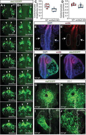
Wnt9b has a permissive role during the anterior convergence movements and fusion of cardiomyocyte progenitor cells. (A,B) Single images from light-sheet microscopy-derived time-lapse movies of cardiomyocyte progenitor cell migrations. Tracks of cardiac progenitor cell migrations during cardiac cone formation are highlighted. In the wildtype (A), cardiomyocyte progenitor cells migrate towards the embryonic midline and fuse first posteriorly and then anteriorly. In wnt9a/b double morphants (B), cardiomyocyte progenitor cells complete migrations and fuse posteriorly, but fail to converge anteriorly. Cardiomyocyte progenitor cells included in the tracking analysis (shown in C,D) have been marked. Dashed yellow lines and white arrowheads indicate the converging anterior part of the heart cone. All the other cell tracks are marked with white dots. (C,D) Tracking analyses of cardiomyocyte progenitor cells during cardiac cone formation. Quantification of the straightness index (C) and velocity (D) of wild-type versus wnt9a/b double morphant cardiomyocyte progenitor cells in anterior positions (wildtype, n=5 embryos; wnt9a/b double morphants, n=5 embryos). Box plots show mean values (middle bars) and first to third interquartile ranges (boxes). Lower and upper whiskers indicate minimum and maximum values, respectively (*P<0.05, **P<0.01; two-tailed, unpaired Student's t-test). (E-F′) Fluorescent whole-mount in situ hybridization of wnt9b expression at stage of cardiomyocyte progenitor cell convergence towards the embryonic midline. (E,E′) The lateral plate mesoderm (white arrows, dashed line) is devoid of wnt9b expression. Instead, wnt9b expression is in the neural tube (white asterisks) and neural retina (n=10/10 embryos analyzed). (F,F′) Neural progenitor cells expressing Tg(elavl3:EGFP)knu3 do not co-express wnt9b (n=10/10 embryos analyzed). (G-J) Maximum projections of confocal z-scan sections with ventral views of cardiomyocyte progenitor cells. In wildtype, cardiomyocyte progenitor cells have fused into the heart cone (G). Upon heat shock-induced overexpression of wnt9b, the size of the heart cone increases (n=22/22 embryos analyzed) (H). At 24 hpf, wild-type cardiomyocyte progenitor cells undergo leftward cardiac jogging (I). Upon heat shock-induced overexpression of wnt9b, cardiomyocyte progenitor cells undergo leftward jogging but are more dispersed (J; n=13/13 embryos analyzed). The embryonic midline is marked by ZO-1 staining (G,H) and dashed lines (I,J). Scale bars: 40 µm (A,B); 30 µm (E-J).
|