FIGURE
Fig. 3
- ID
- ZDB-FIG-230816-9
- Publication
- Légaré et al., 2023 - Loss of mitochondrial Chchd10 or Chchd2 in zebrafish leads to an ALS-like phenotype and Complex I deficiency independent of the mitochondrial integrated stress response
- Other Figures
- All Figure Page
- Back to All Figure Page
Fig. 3
|
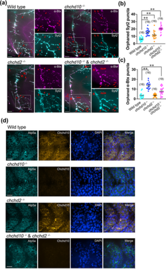
Larval chchd10?/?, chchd2?/?, and double chchd10?/? and chchd2?/? display altered neuromuscular junction (NMJ) integrity, but no obvious mitochondrial network alterations. (a) Representative images of larval ventral trunk NMJs and mitochondrial network in 2 dpf zebrafish. Markers are Syt2 (cyan, presynaptic) and ?Btx (magenta, postsynaptic). Red arrows and red arrowheads in example NMJ images represent orphaned ?Btx and Syt2 puncta, respectively. Scale bars represent 100 ?m. (b) Tabulation of orphaned presynaptic (Syt2) puncta. All genetic groups were significantly different than wild type NMJs (chchd10?/?, p < .0001; chchd2?/?, p = .0281; double chchd10?/? and chchd2?/?, p < .0001). Data shown as mean ± SEM. (c) Tabulation of orphaned postsynaptic (?Btx) puncta. Both chchd10?/? and double chchd10?/? and chchd2?/? showed increased orphaned AChR clusters when compared to wild type larvae (chchd10?/?, p < .0001; double chchd10?/? and chchd2?/?, p = .0069). Data shown as mean ± SEM. Sample sizes (n) are noted in parentheses. Significance was assessed using a one-way ANOVA and Tuckey's multiple comparison test, and significant differences from wild type larvae are represented by either a single asterisk (p < .05) or double asterisk (p < .01). (d) Representative confocal images of the mitochondrial network in the superficially located slow-twitch muscle cell layer. Markers are Atp5a (cyan), DAPI (blue), and Chchd10 (orange). Scale bars represent 40 ?m.
|
Expression Data
Expression Detail
Antibody Labeling
Phenotype Data
Phenotype Detail
Acknowledgments
This image is the copyrighted work of the attributed author or publisher, and
ZFIN has permission only to display this image to its users.
Additional permissions should be obtained from the applicable author or publisher of the image.
Full text @ Dev. Neurobiol.

