
Fig. 3
- ID
- ZDB-FIG-230607-12
- Publication
- Rayrikar et al., 2022 - Ccn2a-FGFR1-SHH signaling is necessary for intervertebral disc homeostasis and regeneration in adult zebrafish
- Other Figures
- All Figure Page
- Back to All Figure Page
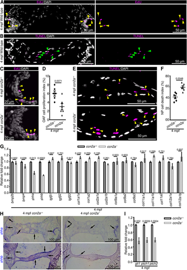
Adult ccn2a−/− display decreased OAF cell proliferation, increased NP cell death and decreased SHH signaling. (A,B) Maximum intensity projections (MIPs) of confocal images of sagittal IVD sections stained for EdU (magenta; proliferating cells) and nuclei (white; DAPI) (A) or stained for TUNEL (magenta; dead cells) and nuclei (white; DAPI) (B). Yellow and green arrowheads indicate EdU+ OAF cells and TUNEL+ NP cells, respectively. (C) MIPs of confocal images of sagittal IVD sections stained for EdU (magenta; proliferating cells) and stained with DAPI (white; nuclei). Arrowheads indicate EdU+ cells. (D) Quantification of OAF cell proliferation (n=6). (E) MIPs of confocal images of IVD sagittal sections stained for TUNEL (magenta; dead cells) and stained with DAPI (white; nuclei). Arrowheads indicate TUNEL+ NP cells. (F) Quantification of NP cell death (n=6). (G) qPCR to identify differentially expressed genes in 4 mpf ccn2a−/− vertebral tissues (n=4). (H) shha and shhb expression on sagittal sections of IVD. Arrows indicate mRNA expression. (I) Quantification of gli1, ptch1 and ptch2 expression in vertebral tissues (n=4). In D,F,G,I, data are mean±s.e.m.; each sample represents one animal. Mean Ct values can be found in Table S5. |