
Fig. 1
- ID
- ZDB-FIG-230607-10
- Publication
- Rayrikar et al., 2022 - Ccn2a-FGFR1-SHH signaling is necessary for intervertebral disc homeostasis and regeneration in adult zebrafish
- Other Figures
- All Figure Page
- Back to All Figure Page
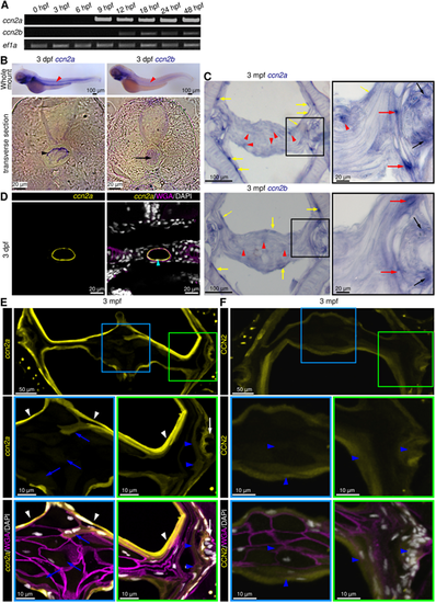
ccn2a and ccn2b are expressed in larval notochord and adult IVDs. (A) Semi-qPCR analysis of ccn2a and ccn2b in zebrafish embryos. ef1a is loading control. (B) Bright-field images of whole-mount in situ hybridized embryos and their transverse sections. Red arrowheads indicate ccn2a or ccn2b expression in the notochord. Black arrowhead and arrow show ccn2a transcripts in the NSCs and ccn2b transcripts in the NCs, respectively. (C) ccn2a and ccn2b expression on sagittal sections of an IVD. Black, red and yellow arrows indicate ccn2a- or ccn2b-expressing cells in the OAF, IAF and NSCs, respectively. Red arrowheads show weak ccn2a and ccn2b expression in the NP cells. (D) BACccn2a:EGFP expression (yellow) in a transverse section of a 3 dpf embryo stained using WGA (magenta; cell membrane) and DAPI (white; nuclei). Arrowhead indicates EGFP expression in the NSCs. (E) BACccn2a:EGFP expression (yellow) in a sagittal section of IVD stained using wheat germ agglutinin (magenta; cell membrane) and DAPI (white; nuclei). White arrows and arrowheads indicate EGFP expression in OAF and sheath cells, respectively. Blue arrows and arrowheads indicate weak EGFP expression in NP and IAF cells, respectively. (F) Maximum intensity projections of confocal images of a sagittal section of IVD immunostained for Ccn2 (yellow), and counterstained using WGA (magenta; cell membrane) and DAPI (white; nuclei). Arrowheads indicate ubiquitous localization of Ccn2 throughout the IVD. |