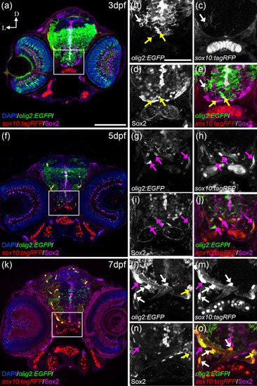
FIGURE 5
- ID
- ZDB-FIG-230420-77
- Publication
- Santos-Ledo et al., 2022 - Oligodendrocyte origin and development in the zebrafish visual system
- Other Figures
- All Figure Page
- Back to All Figure Page
|
olig2:EGFP cells switch from Sox2 to sox10:tagRFP. At 3 dfp (a), olig2:EGFP cells with round morphologies and slightly arborized in the postchiasmatic ON colocalized with Sox2 but not with sox10:tagRFP (a, yellow arrows in b, d, and e). Fully arborized olig2:EGFP cells in the ventral OT colocalized with sox10:tagRFP and not with Sox2 (a, white arrows in b, c, and e). At 5 dpf (f), olig2:EGFP with elongated morphologies and arborization colocalize simultaneously with Sox2 and sox10:tagRFP (f, magenta arrows in g?j). At 7 dfp (k), arborized olig2:EGFP cells that present many projections colocalize only with sox10:tagRFP (k, white arrows in l, m, and o); while some olig2:EGFP cells with less arborization colocalize with Sox2 and sox10:tagRFP (magenta arrows in l?o) and those with round morphologies colocalized only with Sox2 and not sox10:tagRFP (yellow arrows in l, n, and o). D: dorsal; L: lateral. Scale bar in a, f, k: 100 ?m; in b, c, d, e, g, h, I, j, l, m, n, o: 50 ?m |

