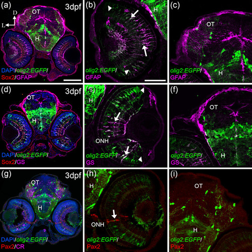
FIGURE 3
- ID
- ZDB-FIG-230420-75
- Publication
- Santos-Ledo et al., 2022 - Oligodendrocyte origin and development in the zebrafish visual system
- Other Figures
- All Figure Page
- Back to All Figure Page
|
olig2:EGFP cells present other glial markers in the retina; GFAP and GS. olig2:EGFP cells also colocalized with Müller markers: GFAP (a, b) and GS (d, e) in the retina but not in the OT (c, f). Colocalization was detected mostly in the central area of the retina (arrows in band e), while cells in the periphery were just olig2:EGFP (arrowheads in b and e). olig2:EGFP cells in the periphery of the ONH do not colocalize with Pax2, typical maker for reticular astrocytes (g?i), although these two populations are very close (arrow in h). D: dorsal; L: lateral; H: hypothalamus; ONH: optic nerve head; OT: optic tectum. Scale bar in a, d, g: 100 ?m; in b, c, e, f, h, i: 50 ?m |

