FIGURE

FIG 4

ID
ZDB-FIG-230420-48
Publication
Nielson et al., 2023 - Roles for Microglia in Cryptococcal Brain Dissemination in the Zebrafish Larva
Other Figures
All Figure Page
Back to All Figure Page
FIG 4

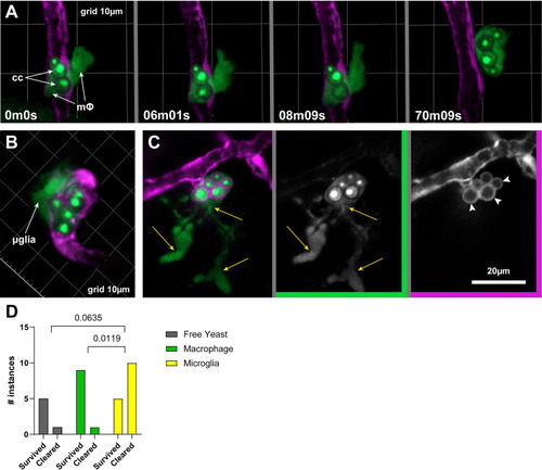
Macrophage and microglia in BBB crossing and subsequent fate of Cryptococcus. (A) Intravascular macrophage (mΦ) containing cryptococcal yeast cells (cc) extends a pseudopod outside the vasculature and pulls its cargo along with it. Phagocytes (mpeg+) and yeast cells express EGFP. Note that nuclear-localized fluorescence signal emphasizes yeast cells versus phagocytes of the same color. Endothelial cells express cytoplasmic mCherry (magenta). (B) A presumed microglial cell (μglia) tightly associated with the abluminal side of a vessel containing Cryptococcus. (C) A cell with microglial morphology in the parenchyma (yellow arrows indicate ramifications) containing yeast cells along with endothelial cytoplasm (white arrowheads). (D) Fate of crossing instances compiled from the original control data set plus additional replicates performed specifically for this analysis. P values indicate results of a Fisher’s exact test applied to contingency data.

Expression Data

Expression Detail
Antibody Labeling
Phenotype Data

Phenotype Detail
Acknowledgments
This image is the copyrighted work of the attributed author or publisher, and ZFIN has permission only to display this image to its users. Additional permissions should be obtained from the applicable author or publisher of the image. Full text @ Microbiol Spectr