FIG 1
- ID
- ZDB-FIG-230420-44
- Publication
- Nielson et al., 2023 - Roles for Microglia in Cryptococcal Brain Dissemination in the Zebrafish Larva
- Other Figures
- All Figure Page
- Back to All Figure Page
|
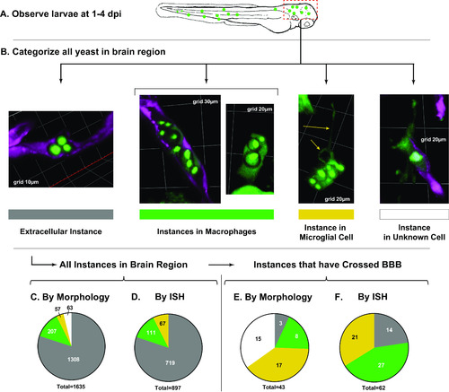
Quantification of cryptococcal yeast cells in the brain from 1 to 4 dpi. (A) Schematic workflow. Instances of cryptococcal yeast in the brain were quantified in observation and enumerated in the brains of a total of 77 live larvae compiled in 3 replicates. (B) Instances were categorized based on morphology. Note that cells with macrophage morphology are seen both inside and outside the vasculature. (C) Morphological quantification of all brain instances in live larvae. Colors correspond with the bars in B; gray, extracellular; green, macrophages; yellow, microglia; white, uncertain morphology. (D) Quantification of all brain instances in a separate set of fixed larvae using ISH for positive identification of microglia. (E) Quantification of the subset of instances that have crossed the BBB in the live larva set. (F) Quantification of the subset of instances that had crossed in the fixed larva set. |
| Fish: | |
|---|---|
| Condition: | |
| Observed In: | |
| Stage: | Day 5 |

