
Fig. 2
- ID
- ZDB-FIG-221123-32
- Publication
- Sarvari et al., 2021 - The E3 ubiquitin-protein ligase Rbx1 regulates cardiac wall morphogenesis in zebrafish
- Other Figures
- All Figure Page
- Back to All Figure Page
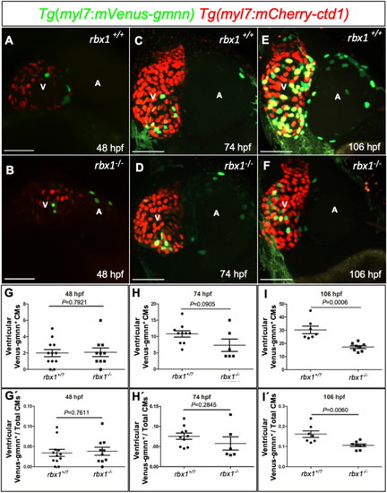
Cardiomyocyte proliferation is reduced in rbx1 mutants (A-F) 3D Confocal images (maximum intensity projections) of Tg(myl7:mVenus-gmnn); Tg(myl7:mCherry-ctd1) hearts at 48 (A?B), 74 (C?D) and 106 (E?F) hpf. (G?I) Quantification of proliferative ventricular CMs from 48 until 106 hpf. At 106 hpf, the number of proliferative CMs in rbx1+/? larvae is significantly higher than that in rbx1?/? siblings (I). (G?-I?) Percentage of proliferative ventricular CMs from 48 until 106 hpf. At 106 hpf, a significantly higher percentage of proliferative CMs is observed in rbx1+/? larvae compared with rbx1?/? siblings (I?). Each dot represents one heart. Data are shown as mean ?� ?SEM. P-values calculated by Student's t-test. V: ventricle, A: atrium; scale bars, 50 ??m. |
| Genes: | |
|---|---|
| Fish: | |
| Anatomical Term: | |
| Stage Range: | Long-pec to Day 4 |
| Fish: | |
|---|---|
| Observed In: | |
| Stage Range: | Long-pec to Day 4 |
Reprinted from Developmental Biology, 480, Sarvari, P., Rasouli, S.J., Allanki, S., Stone, O.A., Sokol, A., Graumann, J., Stainier, D.Y.R., The E3 ubiquitin-protein ligase Rbx1 regulates cardiac wall morphogenesis in zebrafish, 1-12, Copyright (2021) with permission from Elsevier. Full text @ Dev. Biol.