Fig. 1
- ID
- ZDB-FIG-220701-49
- Publication
- Basheer et al., 2022 - In vivo impact of JAK3 A573V mutation revealed using zebrafish
- Other Figures
- All Figure Page
- Back to All Figure Page
|
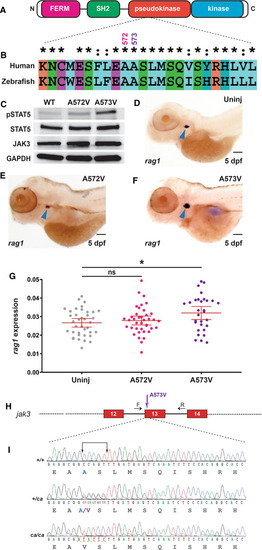
Conservation of human JAK3 constitutively-activating mutations in zebrafish. A JAK3 structure. Schematic representation of JAK3 including the FERM (pink), SH2 (green), pseudokinase (red) and kinase (blue) domains, which are all present in zebrafish Jak3. B Conservation of human and zebrafish JAK3. Human JAK3 and zebrafish Jak3 were aligned using CLUSTALX, showing identical (*) and highly similar (:) residues around A572/A573. C Analysis of zebrafish Jak3 mutants in vitro. Western blot analysis of HEK293 cells transfected with wild-type Jak3?+?Stat5.1 (WT), Jak3 A572V?+?Stat5.1 (A572V) and Jak3 A573V?+?Stat5.1 (A573V) using anti-phospho-STAT5 (pSTAT5), anti-STAT5, anti-HA (JAK3) and anti-GAPDH antibodies, as indicated. D?G Analysis of zebrafish Jak3 mutants in vivo. Uninjected (Uninj) embryos (D) or those injected with mRNA encoding Jak3 A572V (E) or A573V (F), were subjected to WISH with rag1 and imaged, with representative embryos shown. The area of rag1 expression was determined for individual embryos (G) with mean and SEM shown in red and level of statistical significance indicated (*p?<?0.05, ns: not significant; n?=?30) (6.3?×?magnification, scale bar?=?100 ?m). H Genome targeting of zebrafish jak3. Exon?intron structure of the target site with spanning primers (F, R) indicated by black arrows, with exons encoding the pseudokinase domain shown as numbered boxes and introns represented as solid lines, with the Jak3 A573V mutation denoted by a purple arrow. I The jak3 constitutively activating (ca) mutant alleles generated in zebrafish. The nucleotide sequence of homozygote wild-type (+/+), along with heterozygote (+/ca) and homozygote (ca/ca) mutant zebrafish are shown with the five bases mutated indicated and the introduced BciVI site boxed. The protein translations are shown below in black text, with the exception of wild-type A573 in blue and mutant V573 in purple |

