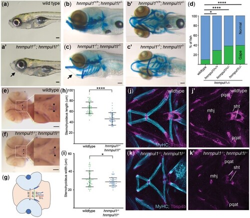
hnrnpul1/1l mutants show a shortened sternohyoideus tendon. a, a’) Images of live 8 dpf wild type and hnrnpul1/1l mutant larvae. b–c’) Lateral (b, c), and ventral (b’, c’) Alcian blue staining at 8 dpf in normal hnrnpul1l−/− mutant (b, b’) and a gaping-jaw phenotype in hnrnpul1/1l mutant (arrows) (c, c’). d) Quantification of the proportion of fish showing a gaping-jaw phenotype. Wild type n = 283, hnrnpul1l−/−; hnrnpul+/+ n = 24, hnrnpul1l−/−; hnrnpul1+/− n = 64, hnrnpul1−/−hnrnpul1l−/− n = 84 from 5 trials. e, f) WISH staining for scleraxis (scxa) in the sternohyoideus tendon (box, arrow heads) in wild type and hnrnpul1/1l mutant embryos at 72 hpf. g) Schematic showing craniofacial tendons and location of tendon measurements. A-W = width between adductor mandibulae tendons, P-W = width between Palatoquadrate tendons, S-W= width between sternohyoideus tendons, S-L = sternohyoideus length. h, i) Quantification of the length (h) and width (i) between the sternohyoideus tendons in wild type (n = 51) and hnrnpul1/1l mutant (n = 50) embryos, from 3 trials. j–k’) Muscle (MyHC, blue) and tendon (Thbs4b, magenta) staining of the craniofacial region of 72 hpf wild type (j, j’) and hnrnpul1/1l mutant (k, k’). *P ≤ 0.05, ****P ≤ 0.0001 determined by Fisher’s exact test (d) and Student’s t-test (h, j) Scale bars = 100 µm. mhj, mandibulohyoid junction; pqat, palatoquadrate adductor tendon; sht, sternohyoideus tendon.
|
