IMAGE

Fig. 6.

ID
ZDB-IMAGE-220509-11
Genes
Antibodies
Source
Figures for Blackwell et al., 2022
Image
Figure Caption

Fig. 6.

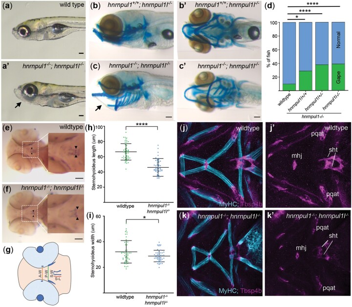
hnrnpul1/1l mutants show a shortened sternohyoideus tendon. a, a’) Images of live 8 dpf wild type and hnrnpul1/1l mutant larvae. b–c’) Lateral (b, c), and ventral (b’, c’) Alcian blue staining at 8 dpf in normal hnrnpul1l−/− mutant (b, b’) and a gaping-jaw phenotype in hnrnpul1/1l mutant (arrows) (c, c’). d) Quantification of the proportion of fish showing a gaping-jaw phenotype. Wild type n = 283, hnrnpul1l−/−; hnrnpul+/+n = 24, hnrnpul1l−/−; hnrnpul1+/−n = 64, hnrnpul1−/−hnrnpul1l−/− n = 84 from 5 trials. e, f) WISH staining for scleraxis (scxa) in the sternohyoideus tendon (box, arrow heads) in wild type and hnrnpul1/1l mutant embryos at 72 hpf. g) Schematic showing craniofacial tendons and location of tendon measurements. A-W = width between adductor mandibulae tendons, P-W = width between Palatoquadrate tendons, S-W= width between sternohyoideus tendons, S-L = sternohyoideus length. h, i) Quantification of the length (h) and width (i) between the sternohyoideus tendons in wild type (n = 51) and hnrnpul1/1l mutant (n = 50) embryos, from 3 trials. j–k’) Muscle (MyHC, blue) and tendon (Thbs4b, magenta) staining of the craniofacial region of 72 hpf wild type (j, j’) and hnrnpul1/1l mutant (k, k’). *P ≤ 0.05, ****P ≤ 0.0001 determined by Fisher’s exact test (d) and Student’s t-test (h, j) Scale bars = 100 µm. mhj, mandibulohyoid junction; pqat, palatoquadrate adductor tendon; sht, sternohyoideus tendon.

Figure Data
Acknowledgments
This image is the copyrighted work of the attributed author or publisher, and ZFIN has permission only to display this image to its users. Additional permissions should be obtained from the applicable author or publisher of the image. Full text @ G3 (Bethesda)