
FIGURE
Fig. 6
- ID
- ZDB-FIG-220203-94
- Publication
- Missinato et al., 2021 - Zebrafish heart regenerates after chemoptogenetic cardiomyocyte depletion
- Other Figures
- All Figure Page
- Back to All Figure Page
Fig. 6
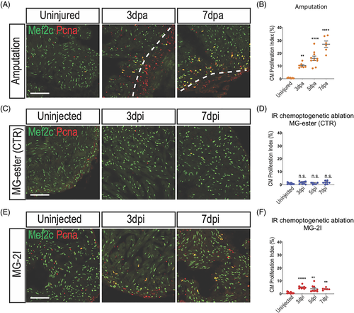
Cardiomyocyte proliferate after chemoptogenetic ablation. A, Mef2c and Pcna staining in amputated ventricles. White dashed lines indicate injury area. Amputation induced cardiomyocyte proliferation. B, Graph showing the percentage of proliferating cardiomyocytes before and after amputation. Cardiomyocyte proliferation started at 3 dpa and peaked at 7 dpa. **P? |
Expression Data
Expression Detail
Antibody Labeling
Phenotype Data
Phenotype Detail
Acknowledgments
This image is the copyrighted work of the attributed author or publisher, and
ZFIN has permission only to display this image to its users.
Additional permissions should be obtained from the applicable author or publisher of the image.
Full text @ Dev. Dyn.