
Fig. 2
- ID
- ZDB-FIG-220203-90
- Publication
- Missinato et al., 2021 - Zebrafish heart regenerates after chemoptogenetic cardiomyocyte depletion
- Other Figures
- All Figure Page
- Back to All Figure Page
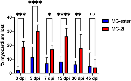
Chemoptogenetic damage causes loss of myocardium after MG-2I treatment. Regions of Interest were taken from WT uninjured, Tg(myl7:fapdl1-cerulean) MG-ester-, and Tg(myl7:fapdl1-cerulean) MG-2I-treated hearts after AFOG staining and intact myocardium was measured using Threshold Particle Analysis (Image J). Myocardial area from MG-ester and MG-2I hearts was normalized to WT uninjured hearts to generate the amount of tissue lost as a percentage. MG-2I-treated hearts showed significant loss of myocardium at 3 dpi and 5 dpi (compared to MG-ester controls), and also regenerated this damage by 45-60dpi. A minimum of three hearts were used per condition with the following as the total n values: WT uninjured (n = 6), MG-ester (3 dpi (n = 5), ?5 dpi (n = 5), ?7 dpi (n = 5), ?15 dpi (n = 5), ?30 dpi (n = 5), ?45 dpi (n = 3)), and MG-2I (3 dpi (n = 5), ?5 dpi (n = 5), ?7 dpi (n = 6), ?15 dpi (n = 6), ?30 dpi (n = 5), ?45 dpi (n = 3)). *P? |