
Fig. 3
- ID
- ZDB-FIG-210405-24
- Publication
- Farrugia et al., 2020 - Mechanisms of vascular damage by systemic dissemination of the oral pathogen Porphyromonas gingivalis
- Other Figures
- All Figure Page
- Back to All Figure Page
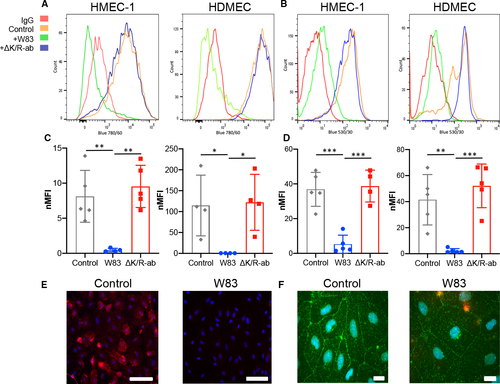
Degradation of endothelial cell surface?expressed junctional adhesion molecules in vitro. Representative flow cytometry histograms of cell surface expression of PECAM?1 (A) and VE?cadherin (B) on HMEC?1 and HDMEC following infection with Pg W83 (green) or ?K/R?ab mutant (blue); IgG control (red); and uninfected control (orange). Normalised median fluorescence intensity (nMFI) histograms of PECAM?1 (C) and VE?cadherin (D) on HMEC?1 and HDMEC following infection with Pg W83, ?K/R?ab or uninfected control (enclosed circles represent data from each individual experiment, n = 4). Statistical differences were analysed by one?way ANOVA with Tukey?s multiple comparison test *P < 0.05, **P < 0.01, ***P < 0.001. Micrograph images show immunofluorescent detection of cell surface expression of PECAM?1 (red, E) and VE?cadherin (green, F) in control or Pg?treated HDMEC (n = 3). Nuclei were counterstained blue with DAPI. Scale bars in E & F = 100 ?m. |