
Fig. 1
- ID
- ZDB-FIG-210405-22
- Publication
- Farrugia et al., 2020 - Mechanisms of vascular damage by systemic dissemination of the oral pathogen Porphyromonas gingivalis
- Other Figures
- All Figure Page
- Back to All Figure Page
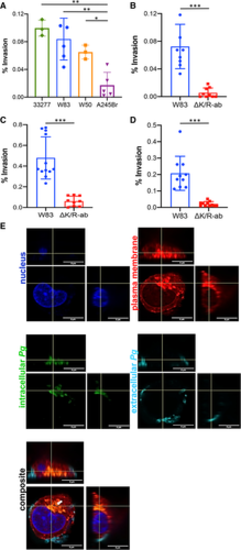
Strain and gingipain?dependent invasion of Pg into human endothelial cells. HDMEC were infected with wild?type strain Pg ATCC33277, W83, W50 or A245Br (A) or wild?type Pg W83 and ?K/R?ab (B) at a MOI 100 for 90 min. HMEC?1 (C) and HCAEC (D) were infected with wild?type Pg W83 or ?K/R?ab mutant also at a MOI 100 for 90 min. Bacterial cell invasion was determined by antibiotic protection assay and expressed as a percentage of primary bacterial inoculum recovered. Graphs show means � SD (n = at least 3 individual experiments with each individual experiment performed in triplicate technical repeats; shown as filled circles). Data were analysed using one?way ANOVA with Tukey?s post hoc comparison test (Fig. 1A) or Student's t?test (Fig 1B?D), *P < 0.05, **P < 0.01, ***P < 0.001. Representative maximum intensity Z projection images of HMEC?1 with intracellular (green) and extracellular (cyan) Pg W83. Cell nucleus (blue) and plasma membrane (red). Composite image shows intracellular dwelling Pg as orange (green and red colocalisation, white arrow), and extracellular (cyan) bacteria bound to the cell surface (E). All images show x?axis, y?axis and z?axis planes, and scale bars in images are all 5 ?m. |