|
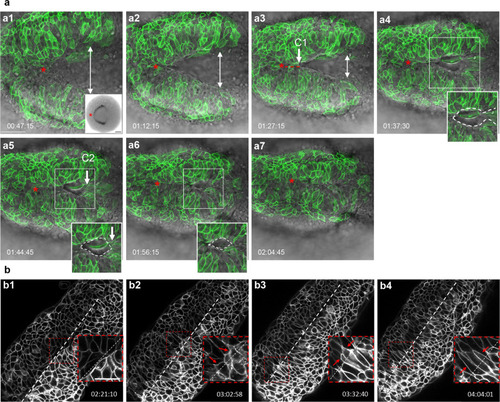
Dynamics of neural fold fusion.a, a1–a7 Time-lapse movie frames of an embryo expressing mosaic GFP, imaged from a dorsal view, showing the initiation of neural tube closure. Images are overlays of the green and brightfield channels. Inset in a1 shows a dorsal view of an emx3-labeled embryo. Insets in a4–a6 outline the eye-shaped opening that forms between closure sites one C1 and two (C2). b Greyscale time-lapse movie frames of an mGFP-labeled embryo imaged from a dorsal view, revealing the final stages of neural fold fusion. Insets in the lower right corner of b1–b4 are higher magnification views of boxed areas. C1, C2 closure sites one and two. Annotations: red asterisk: apex of the neural fold arc; red dotted line: synchronous and posteriorly-directed neural fold fusion anterior to closure point one; white double arrows: distance between the neural folds; white dotted oval: eye-shaped opening, the corners of which are defined by closure points one and two; white dotted line: embryonic midline; red arrows: filopodia extending across the midline; time-elapsed is shown at the bottom of each panel. Scale bars: 50 μm in a1, 100 μm in a1 inset, and 25 μm in b1.
|