FIGURE

Fig 2

ID
ZDB-FIG-210128-23
Publication
Petratou et al., 2021 - The MITF paralog tfec is required in neural crest development for fate specification of the iridophore lineage from a multipotent pigment cell progenitor
Other Figures
All Figure Page
Back to All Figure Page
Fig 2

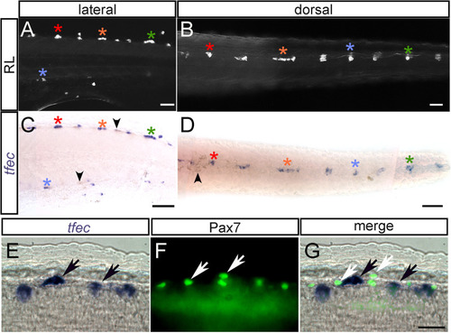
<italic>tfec</italic> is a marker of differentiated iridophores, but not xanthophores.

(A–D) Chromogenic whole-mount in situ hybridisation reveals tfec expression in iridophores at 3 dpf. Iridophores (asterisks) of the posterior trunk/anterior tail are imaged live with reflected light in lateral (A) and dorsal (B) views. (C,D) Whole-mount in situ hybridisation on the same embryo reveals a pattern of tfec expression matching that of the differentiated iridophores (asterisks). The colour of the asterisks indicates corresponding iridophore patches between A and C, and B and D. Note the absence of expression in the location of the associated melanocytes (residual melanin indicated by arrowheads) (E-G). Expression of tfec (E, in situ hybridisation) and of Pax7 (F, immunofluorescence) in differentiated iridophores and xanthophores, respectively, at 48 hpf; note the absence of co-expression of these markers. A,C,E: lateral views; B,D: dorsal views. Head towards the left. Scale bars: A-D, 50 μm; E-G, 20 μm.

Expression Data
Gene:
Antibody:
Fish:
Anatomical Terms:
Stage Range: Long-pec to Protruding-mouth

Expression Detail
Antibody Labeling
Phenotype Data

Phenotype Detail
Acknowledgments
This image is the copyrighted work of the attributed author or publisher, and ZFIN has permission only to display this image to its users. Additional permissions should be obtained from the applicable author or publisher of the image. Full text @ PLoS One