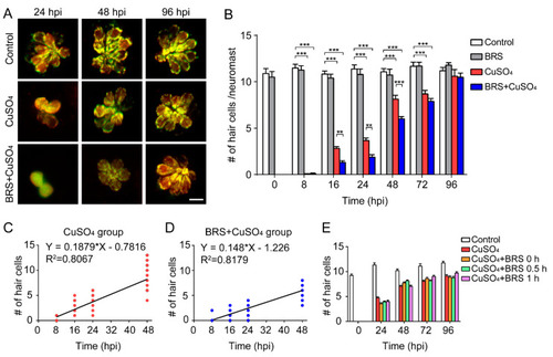
Suppressing inflammation delays hair cell regeneration. (A) Real-time imaging (40×) displays regenerated hair cells in the CuSO4 and BRS+CuSO4 group at 24, 48, and 96 hpi. The control group was imaged at the same time points. Scale bar represents 10 μm. (B) The numbers of regenerated hair cells were significantly lower in the BRS+CuSO4 group compared to the CuSO4 group at 16 (n ≥ 27, p < 0.01), 24 (n ≥ 21, p < 0.01), and 48 (n ≥ 24, p < 0.001) hpi. At 96 hpi, hair cells in both the CuSO4 group and the BRS+CuSO4 group had regenerated to normal levels. Linear analysis in the CuSO4 group (C) and BRS+CuSO4 group (D) was conducted on the number of regenerated cells within 48 hpi. The slope in the CuSO4 group (0.188) was higher than that in the BRS+CuSO4 group (0.148), and the x-intercept in the CuSO4 group (4.160) is higher than that in the BRS+CuSO4 group (8.287). (E) When the time window of inflammatory suppression was delayed, there was no delay in the regeneration of hair cells. BRS-28 was added at the same time as CuSO4 (CuSO4+BRS 0 h group), 30 min after the addition of CuSO4 (CuSO4+BRS 0.5 h group), or 1 h after the addition of CuSO4 (CuSO4+BRS 1 h group) (n ≥ 27 neuromasts at each time point of each group). For (B) and (E), comparisons were performed using two-way ANOVA, with Tukey’ multiple comparisons test. All error bars show the mean ± S.E.M., *** p < 0.001, ** p < 0.01.
|

