FIGURE

Figure 4—figure supplement 2.

ID
ZDB-FIG-200822-23
Publication
Yang et al., 2020 - Drainage of inflammatory macromolecules from brain to periphery targets the liver for macrophage infiltration
Other Figures
All Figure Page
Back to All Figure Page
Figure 4—figure supplement 2.

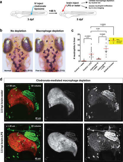
Clodronate-mediated macrophage depletion results in an increase in neutrophil infiltration after brain-LPS injection.

(a) Schematic illustrating the method. Intravenous injection of clodronate liposome was performed at three dpf to allow time for the clodronate to effectively induce cell death to most macrophages over a 48 hr period. (b) Assessment of macrophage depletion by neutral red staining shows that most animals injected with clodronate liposome indeed resulted in an apparent loss of macrophages in the brain (58%, n = 12). Arrows point to microglia in the ‘no depletion’ category. (c) Scatter plot shows number of neutrophils infiltrating the liver to be even more increased after macrophage ablation in response to brain-LPS injection. Since clodronate based macrophage depletion is effective only in a proportion of the animals injected, we also used the cluster of the largest numbers of infiltrating neutrophils as a subset to show the animals with the most significant increase. (d) 3D volumetric view of the whole liver from imaging transgenic reporters fabp10a:DsRed for hepatocytes and lyz:GFP for neutrophils. Left, merged channels. Middle, DsRed channel. Right, GFP channel. Brain-water sample is shown 60 µm beneath the first surface of the liver, and the brain-LPS sample is 45 µm below the most outer liver surface. Significantly higher number of neutrophils (arrows) is easily visualized in the brain-LPS sample. Dextran-Alexa 488 was co-injected into the brain as a tracer to validate injections, and can be seen labeling the pronephros in the GFP channel. Statistical significance was determined by a two-tailed t-test and with Welch’s correction for unequal variances as determined by a F-test.

Expression Data

Expression Detail
Antibody Labeling
Phenotype Data

Phenotype Detail
Acknowledgments
This image is the copyrighted work of the attributed author or publisher, and ZFIN has permission only to display this image to its users. Additional permissions should be obtained from the applicable author or publisher of the image. Full text @ Elife