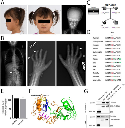
Identification of a de novo mutation in human PHETA1. (A) Images of the UDP patient, presenting with facial asymmetry, concave nasal ridge and malar flattening. Radiograph (right) reveals mild asymmetry of the skull. (B) Radiographs depict scoliosis (arrowhead in left image) and clinodactyly of fourth and fifth digits on both hands (arrowheads in middle and right images). (C) Whole-exome sequencing was performed on both parents and the fraternal twin of the UDP patient. ‘N’ denotes not affected and ‘Y’ denotes affected. The arrow labeled ‘P’ identifies the UDP patient (UDP_5532). ‘+’ indicates the presence of a normal allele, thus marking p.R6C as a heterozygous mutation. (D) COBALT multiple alignment of partial protein sequences of PHETA1 orthologs. The conserved arginine residue is highlighted in red, and amino acid residues that differ from the sequence of the human PHETA1 protein are highlighted in green. The arginine residue is highly conserved across multiple species. (E) Relative quantification of mRNA expression in the patient cells showing that the expression of PHETA1 is not significantly different from that in controls. Error bar represents s.d. from six replicates. (F) 3-D structure of the human PHETA1 protein showing the PH domain (green) with a four-stranded N-terminal and three-stranded C-terminal β-sheet with a helix (orange). The conserved arginine amino acid (Arg19 in the PHETA1 long isoform, yellow) is far from the F&H motif (magenta); however, it stabilizes the folded domain around the C-terminal helix. (G) GFP-tagged full-length WT PHETA1 or PHETA1R6C were expressed in HeLa cells and tested for interaction with full-length HA-tagged OCRL1. Bound proteins detected by western blotting with the indicated antibodies. ‘IP: anti-GFP’ refers to the anti-GFP antibody-bound fraction; ‘Total’ represents the total cell lysate.
|