
FIGURE
Fig. 4
- ID
- ZDB-FIG-200514-44
- Publication
- Paatero et al., 2018 - Junction-based lamellipodia drive endothelial cell rearrangements in vivo via a VE-cadherin-F-actin based oscillatory cell-cell interaction
- Other Figures
- All Figure Page
- Back to All Figure Page
Fig. 4
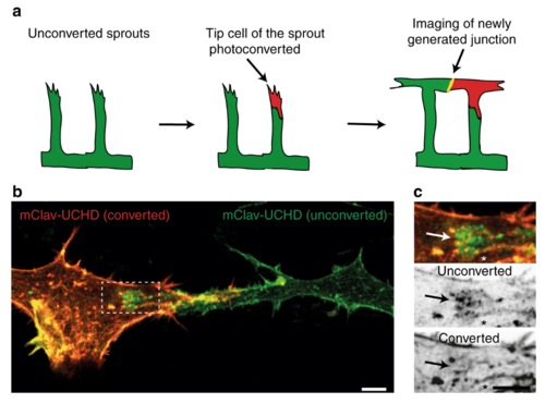
JBL formation at the distal tip of the junction during DLAV anastomosis. a Schematic representation of the mClav2-UCHD photoconversion experiment. b Image of photoconverted and unconverted mClav2-UCHD cells in the DLAV of an Tg(fli:Gal4ffubs3;UAS:mClav2-UCHDubs27) embryo, at 32?hpf. c A close up of the inset in b. Arrows point to differentially labeled JBL and asterisk (*) marks the junction outside JBL. Scale bar 5?�m |
Expression Data
| Gene: | |
|---|---|
| Fish: | |
| Anatomical Terms: | |
| Stage: | Prim-15 |
Expression Detail
Antibody Labeling
Phenotype Data
Phenotype Detail
Acknowledgments
This image is the copyrighted work of the attributed author or publisher, and
ZFIN has permission only to display this image to its users.
Additional permissions should be obtained from the applicable author or publisher of the image.
Full text @ Nat. Commun.