|
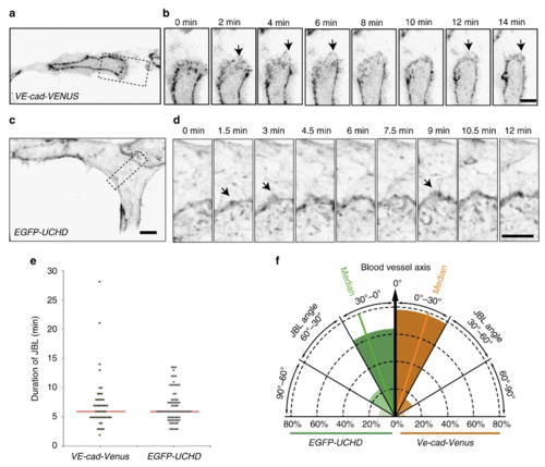
Active VE-cad and F-actin behavior of junction-based lamellipodia. a, b Still images from a movie (Supplementary Movie 2) of a VE-cad-Venus expressing embryo Tg(BAC(cdh5:cdh5-ts)), showing the DLAV at 30?hpf in inversed contrast. b A magnification of the inset in a. Arrows point to JBL. c, d Still images from a movie (Supplementary Movie 3) of a EGFP-UCHD expressing embryo (Tg(fli:Gal4ffubs3, UAS:EGFP-UCHDubs18)) showing the DLAV at 30?hpf in inversed contrast. d A magnification of the inset in c. Arrows point to JBL. e Scatter plot of quantitation of the duration of the JBL with the VE-cad-Venus transgene (n?=?48 in 6 embryos) and EGFP-UCHD movies (n?=?74 in 6 embryos), red line represents the median. f Quantitation of JBL angle in the DLAV in respect to the blood vessel axis (0�) using the EGFP-UCHD transgene (n?=?103 from 6 embryos) or Cdh5-Venus transgene (n?=?41 from 5 embryos). Scale bars 5?�m
|