
Fig. 7
- ID
- ZDB-FIG-200306-122
- Publication
- Nimura et al., 2019 - Role of Reelin in cell positioning in the cerebellum and the cerebellum-like structure in zebrafish
- Other Figures
- All Figure Page
- Back to All Figure Page
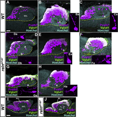
Defects in the migration and polarity of PCs in reln mutants. Sagittal sections of the brain from WT (A-C, n=?4) and reln?7/?7 (D-H, n=?4) 15-dpf larvae, and from WT (I, n=?3) and reln?7/?7 (J, n=?3) 30-dpf fish, were stained with anti-Pvalb7 (magenta) and anti-Vglut1 (green) antibodies, and Hoechst (cyan). Typical cerebellum images are shown. The ventral limit of the cerebellum is indicated by a dotted line. (Ba, Ca, Da, Ea, Fa, Ga, Ha) High magnification images of the PCs marked by ?a? in B?H. The Pvalb7 and Vglut1-double positive region marks the ML. Migrating PCs were detected in the GCL in WT, and they extended a neurite (primary dendrite) toward the pial side (Ba, Ca, n=?4). Many of the migrating cells extended one or multiple neurites in aberrant directions in the reln?7/?7mutants (Ea, Fa, Ga, Ha, n=?4). At 30 dpf, most of the PCs had reached the PCL in WT (I), whereas many ectopic PCs were detected in the GCL in reln?7/?7 mutants (J, n=?3). The abbreviations are described in the legend for Fig. 1. Scale bars: 20??m in A (applies to A-H); 100??m in I (applies to I-J). |
| Genes: | |
|---|---|
| Antibodies: | |
| Fish: | |
| Anatomical Terms: | |
| Stage Range: | Days 14-20 to Days 30-44 |
| Fish: | |
|---|---|
| Observed In: | |
| Stage Range: | Days 14-20 to Days 30-44 |
Reprinted from Developmental Biology, 455(2), Nimura, T., Itoh, T., Hagio, H., Hayashi, T., Di Donato, V., Takeuchi, M., Itoh, T., Inoguchi, F., Sato, Y., Yamamoto, N., Katsuyama, Y., Del Bene, F., Shimizu, T., Hibi, M., Role of Reelin in cell positioning in the cerebellum and the cerebellum-like structure in zebrafish, 393-408, Copyright (2019) with permission from Elsevier. Full text @ Dev. Biol.